ARG70235
Human CD279 / PD-1 recombinant protein (His-tagged, C-ter)
Human CD279 / PD-1 recombinant protein (His-tagged, C-ter) for Binding Activity,SDS-PAGE and Human
Overview
| Product Description | HEK293 expressed, His-tagged (C-ter) Human CD279 / PD-1 recombinant protein. |
|---|---|
| Tested Reactivity | Hu |
| Tested Application | Binding, SDS-PAGE |
| Target Name | CD279 / PD-1 |
| Species | Human |
| A.A. Sequence | Leu25 - Thr168 of Human CD279 / PD - 1 (NP_005009.2) with 6X His tag at the C - terminus. |
| Expression System | HEK293 |
| Alternate Names | hPD-l; CD279; PD-1; Protein PD-1; CD antigen CD279; PD1; hSLE1; SLEB2; Programmed cell death protein 1; hPD-1 |
Application Instructions
| Application Note | Binding activity test: Measured by its binding ability in a functional ELISA. Immobilized Recombinant Human PD-1 at 5 µg/ml (100µl/well) can bind Recombinant Human PD-L1 with a linear range of 0.5-2 µg/ml. |
|---|
Properties
| Form | Powder |
|---|---|
| Purification Note | 0.22 µm filter sterilized. Endotoxin level is <0.1 EU/µg of the protein, as determined by the LAL test. |
| Purity | > 97% (by SDS-PAGE) |
| Buffer | PBS (pH 7.4) |
| Reconstitution | Reconstitute to a concentration of 0.1 - 0.5 mg/ml in sterile distilled water. |
| Storage Instruction | For long term, lyophilized protein should be stored at -20°C or -80°C. After reconstitution, aliquot and store at -20°C for up to one month, at 2-8°C for up to one week. Storage in frost free freezers is not recommended. Avoid repeated freeze/thaw cycles. Suggest spin the vial prior to opening. |
| Note | For laboratory research only, not for drug, diagnostic or other use. |
Bioinformation
| Gene Symbol | PDCD1 |
|---|---|
| Gene Full Name | programmed cell death 1 |
| Background | This gene encodes a cell surface membrane protein of the immunoglobulin superfamily. This protein is expressed in pro-B-cells and is thought to play a role in their differentiation. In mice, expression of this gene is induced in the thymus when anti-CD3 antibodies are injected and large numbers of thymocytes undergo apoptosis. Mice deficient for this gene bred on a BALB/c background developed dilated cardiomyopathy and died from congestive heart failure. These studies suggest that this gene product may also be important in T cell function and contribute to the prevention of autoimmune diseases. [provided by RefSeq, Jul 2008] |
| Function | Inhibitory receptor on antigen activated T-cells that plays a critical role in induction and maintenance of immune tolerance to self (PubMed:21276005). Delivers inhibitory signals upon binding to ligands CD274/PDCD1L1 and CD273/PDCD1LG2 (PubMed:21276005). Following T-cell receptor (TCR) engagement, PDCD1 associates with CD3-TCR in the immunological synapse and directly inhibits T-cell activation (By similarity). Suppresses T-cell activation through the recruitment of PTPN11/SHP-2: following ligand-binding, PDCD1 is phosphorylated within the ITSM motif, leading to the recruitment of the protein tyrosine phosphatase PTPN11/SHP-2 that mediates dephosphorylation of key TCR proximal signaling molecules, such as ZAP70, PRKCQ/PKCtheta and CD247/CD3zeta (By similarity). The PDCD1-mediated inhibitory pathway is exploited by tumors to attenuate anti-tumor immunity and escape destruction by the immune system, thereby facilitating tumor survival (PubMed:28951311). The interaction with CD274/PDCD1L1 inhibits cytotoxic T lymphocytes (CTLs) effector function (PubMed:28951311). The blockage of the PDCD1-mediated pathway results in the reversal of the exhausted T-cell phenotype and the normalization of the anti-tumor response, providing a rationale for cancer immunotherapy (PubMed:22658127, PubMed:25034862, PubMed:25399552). [UniProt] |
| Cellular Localization | Membrane; Single-pass type I membrane protein. [UniProt] |
| Calculated MW | 32 kDa |
Images (1) Click the Picture to Zoom In
-

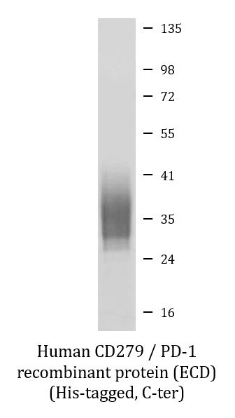
ARG70235 Human CD279 / PD-1 recombinant protein (ECD) (His-tagged, C-ter) SDS-PAGE image
SDS-PAGE analysis of ARG70235 Human CD279 / PD-1 recombinant protein (ECD) (His-tagged, C-ter).