ARG30144
Phospho beta Catenin Antibody Panel (Total, pS33, pS37, pT41 / pS45)
Cancer antibody; Cell Biology and Cellular Response antibody; Developmental Biology antibody; Immune System antibody; Neuroscience antibody; Signaling Transduction antibody
Component
| Cat No | Component Name | Host clonality | Reactivity | Application | Package |
|---|---|---|---|---|---|
| ARG52651 | anti-beta Catenin antibody | Rabbit pAb | Hu, Ms, Rat | ICC/IF, IHC-P, IHC-Fr, IP, WB | 50 μl |
| ARG51709 | anti-beta Catenin phospho (Ser33) antibody | Rabbit pAb | Hu, Ms, Rat | IHC-P, WB | 50 μl |
| ARG51710 | anti-beta Catenin phospho (Ser37) antibody | Rabbit pAb | Hu, Ms, Rat | IHC-P, WB | 50 μl |
| ARG51618 | anti-beta Catenin phospho (Thr41 / Ser45) antibody | Rabbit pAb | Hu, Ms, Rat | WB | 50 μl |
| ARG65351 | Goat anti-Rabbit IgG antibody (HRP) | Goat pAb | Rb | ELISA, IHC-P, WB | 50 μl |
Overview
| Product Description | β-Catenin is a key downstream effector in the Wnt signaling pathway. It is implicated in two major biological processes in vertebrates: early embryonic development and tumorigenesis. CK1 phosphorylates β-catenin at Ser45. This phosphorylation event primes β-catenin for subsequent phosphorylation by GSK-3β. GSK-3β destabilizes β-catenin by phosphorylating it at Ser33, Ser37, and Thr41. Both Akt and PKA were shown to phosphorylate β-catenin at Ser552 and Ser675. Phosphorylation at Ser552 and Ser675 induces β-catenin accumulation in the nucleus and increases its transcriptional activity.This antibody panel investigates different processes through the phosphorylation on multiple site of β-Catenin. Cadigan, K.M. and Nusse, R. (1997) Genes Dev 11, 3286-305. Amit, S. et al. (2002) Genes Dev 16, 1066-76. Liu, C. et al. (2002) Cell 108, 837-47. Yanagawa, S. et al. (2002) EMBO J 21, 1733-42. Yost, C. et al. (1996) Genes Dev 10, 1443-54. Fang D et al. (2007) J Biol Chem 282, 11221–9. He, X.C. et al. (2007) Nat. Genet. 39, 189-198. |
|---|---|
| Target Name | beta Catenin |
| Alternate Names | Phospho beta Catenin antibody; beta Catenin phospho (Thr41 / Ser45) antibody; beta Catenin phospho (Ser33) antibody; beta Catenin phospho (Ser37) antibody; beta Catenin antibody |
Properties
| Storage Instruction | For continuous use, store undiluted antibody at 2-8°C for up to a week. For long-term storage, aliquot and store at -20°C or below. Storage in frost free freezers is not recommended. Avoid repeated freeze/thaw cycles. Suggest spin the vial prior to opening. The antibody solution should be gently mixed before use. |
|---|---|
| Note | For laboratory research only, not for drug, diagnostic or other use. |
Bioinformation
| Gene Full Name | Antibody Panel for Phospho beta Catenin (Total, pS33, pS37, pT41/pS45) |
|---|---|
| Highlight | Related Product: anti-beta Catenin antibody; |
| Research Area | Cancer antibody; Cell Biology and Cellular Response antibody; Developmental Biology antibody; Immune System antibody; Neuroscience antibody; Signaling Transduction antibody |
Images (11) Click the Picture to Zoom In
-

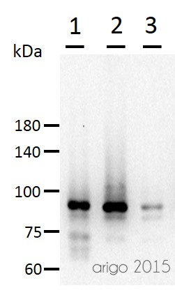
ARG52651 anti-beta Catenin antibody WB image
Western blot: 30 µg of 1) 293T, 2) 3T3, and 3) Mouse liver lysate stained with ARG52651 anti-beta Catenin antibody at 1:500 dilution.
-

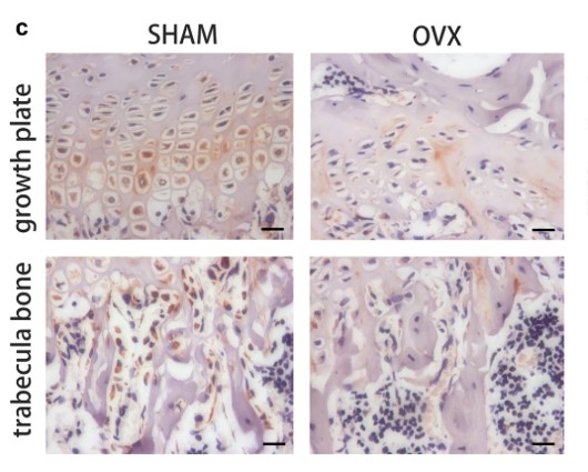
ARG65351 Goat anti-Rabbit IgG antibody (HRP) IHC-P image
From Yu-Qian Song et al. J Mol Med (Berl) (2022), doi: 10.1007/s00109-021-02165-0, Fig. 5.c.
-
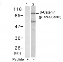
ARG51618 anti-beta Catenin phospho (Thr41/Ser45) antibody WB image
Western Blot: extracts from SW626 cells stained with anti-beta Catenin (phospho Thr41/Ser45) antibody ARG51618 (Lane 2) and the same antibody preincubated with blocking peptide (Lane1).
-
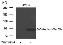
ARG51709 anti-beta Catenin phospho (Ser33) antibody WB image
Western Blot: extracts from MCF-7 cells untreated or treated with Calyculin A stained with anti-beta Catenin (phospho Ser33) antibody ARG51709.
-
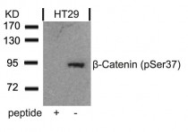
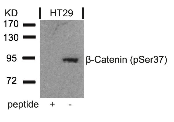
ARG51710 anti-beta Catenin phospho (Ser37) antibody WB image
Western Blot: extracts from HT29 cells stained with anti-beta Catenin (phospho Ser37) antibody ARG51710 and the same antibody preincubated with blocking peptide .
-
ARG52651 anti-Catenin-beta antibody WB image
Western Blot: A431 stained with Catenin-beta antibody (ARG52651)
-
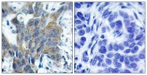
ARG51709 anti-beta Catenin phospho (Ser33) antibody IHC-P image
Immunohistochemistry: paraffin-embedded human breast carcinoma tissue stained with anti-beta Catenin (phospho Ser33) antibody ARG51709 (left) or the same antibody preincubated with blocking peptide (right).
-
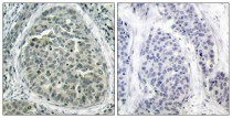
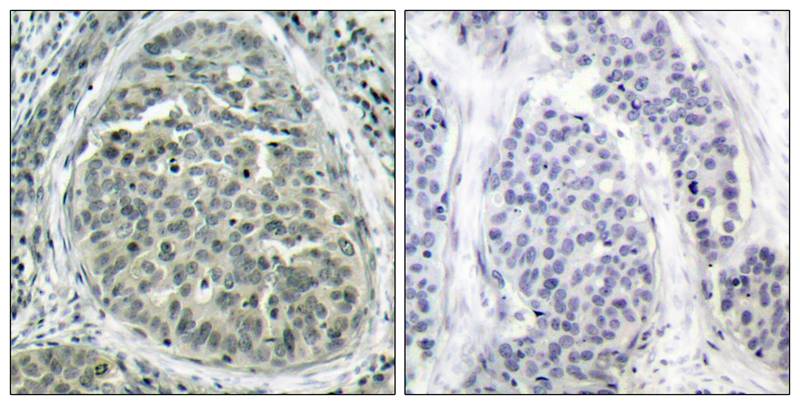
ARG51710 anti-beta Catenin phospho (Ser37) antibody IHC-P image
Immunohistochemistry: paraffin-embedded human breast carcinoma tissue stained with anti-beta Catenin (phospho Ser37) antibody ARG51710 (left) or the same antibody preincubated with blocking peptide (right).
-
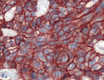
ARG52651 anti-Catenin-beta antibody IHC-P image
Immunohistochemistry: Human Breast Carcinoma stained with Catenin-beta antibody (ARG52651)
-
ARG65351 Goat anti-Rabbit IgG antibody (HRP) WB image
Western blot: Rat placental stained with ARG57589 anti-MTNR1A antibody at 1:1000 dilution, ARG65351 Goat anti-Rabbit IgG antibody (HRP) at 1:5000 dilution.
From Jinzhi Li et al. J Reprod Immunol. (2023), doi: 10.1016/j.jri.2023.104166, Fig. 2.B.
-
ARG65351 Goat anti-Rabbit IgG antibody (HRP) WB image
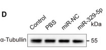
Western blot: Mouse retina stained with ARG65693 anti-alpha Tubulin antibody and ARG65351 Goat anti-Rabbit IgG antibody (HRP)
From Xiaoyuan Ye et al. Mol Ther Nucleic Acids. (2024), doi: 10.1016/j.omtn.2024.102209, Fig. 5.D.


Specific References
Progressively diminished estrogen signaling concordant with increased fibrosis in ectopic endometrium
ARG65351; WB /


The Therapeutic Potential of Exosomes vs. Matrix-Bound Nanovesicles from Human Umbilical Cord Mesenchymal Stromal Cells in Osteoarthritis Treatment
ARG65351; WB /
Environmental acidification drives inter-organ energy mobilization to enhance reproductive performance in medaka (Oryzias latipes)
ARG65351; WB /
Diuretic Action of Apelin-13 Mediated by Inhibiting cAMP/PKA/sPRR Pathway
ARG52651: WB / Mouse
Narasin inhibits tumor metastasis and growth of ERα‑positive breast cancer cells by inactivation of the TGF‑β/SMAD3 and IL‑6/STAT3 signaling pathways
ARG52651: WB / Human
Ubiquitin specific peptidase 5 enhances STAT3 signaling and promotes migration and invasion in Pancreatic Cancer.
ARG52651: WB / Human


