ARG57237
anti-5-hydroxymethylcytosine / 5-hmC antibody [RM236]
anti-5-hydroxymethylcytosine / 5-hmC antibody [RM236] for Dot blot,ELISA,ICC/IF,IHC-Formalin-fixed paraffin-embedded sections,MeDIP and Other
Overview
| Product Description | Rabbit Monoclonal antibody [RM236] recognizes 5-hydroxymethylcytosine / 5-hmC |
|---|---|
| Tested Reactivity | Other |
| Tested Application | Dot, ELISA, ICC/IF, IHC-P, MeDIP |
| Specificity | This antibody reacts to 5-hydroxymethylcytosine in both single-stranded and double-stranded DNA. No cross reactivity with non-methylated cytosine and methylcytosine in DNA. |
| Host | Rabbit |
| Clonality | Monoclonal |
| Clone | RM236 |
| Isotype | IgG |
| Target Name | 5-hydroxymethylcytosine / 5-hmC |
| Antigen Species | Others |
| Immunogen | BSA-conjugated 5-hydroxymethylcytosine. |
| Conjugation | Un-conjugated |
Application Instructions
| Application Suggestion |
|
||||||||||||
|---|---|---|---|---|---|---|---|---|---|---|---|---|---|
| Application Note | * The dilutions indicate recommended starting dilutions and the optimal dilutions or concentrations should be determined by the scientist. |
Properties
| Form | Liquid |
|---|---|
| Purification | Purification with Protein A. |
| Buffer | PBS, 0.09% Sodium azide, 50% Glycerol and 1% BSA. |
| Preservative | 0.09% Sodium azide |
| Stabilizer | 50% Glycerol and 1% BSA |
| Concentration | 1 mg/ml |
| Storage Instruction | For continuous use, store undiluted antibody at 2-8°C for up to a week. For long-term storage, aliquot and store at -20°C. Storage in frost free freezers is not recommended. Avoid repeated freeze/thaw cycles. Suggest spin the vial prior to opening. The antibody solution should be gently mixed before use. |
| Note | For laboratory research only, not for drug, diagnostic or other use. |
Images (6) Click the Picture to Zoom In
-
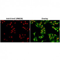
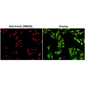
ARG57237 anti-5-hydroxymethylcytosine / 5-hmC antibody [RM236] ICC/IF image
Immunofluorescence: HeLa cells stained with ARG57237 anti-5-hydroxymethylcytosine / 5-hmC antibody [RM236] at 0.5 µg/ml (red). Actin filaments was labeled with fluorescein phalloidin (green).
Cells were fixed with 4% parafor-maldehyde and permeabilized with methanol (-20°C) before treatment with 2 N HCl for 30 min at 37°C to denature DNA.
-
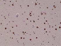
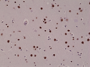
ARG57237 anti-5-hydroxymethylcytosine / 5-hmC antibody [RM236] IHC-P image
Immunohistochemistry: Formalin-fixed and paraffin-embedded Human brain tissue sections stained with ARG57237 anti-5-hydroxymethylcytosine / 5-hmC antibody [RM236].
-
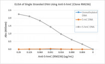
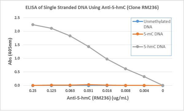
ARG57237 anti-5-hydroxymethylcytosine / 5-hmC antibody [RM236] ELISA image
ELISA: Titration curve of ARG57237 anti-5-hydroxymethylcytosine / 5-hmC antibody [RM236]. Antigen: The plate was coated with streptavidin and then biotinylated single stranded unmethylated DNA, 5-Methylcytosine (5-mC) DNA, and 5-Hydroxymethylcytosine (5-hmC) DNA.
Secondary antibody: An alkaline phosphatase conjugated anti-rabbit IgG.
-
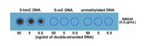
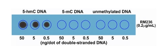
ARG57237 anti-5-hydroxymethylcytosine / 5-hmC antibody [RM236] Dot blot image
Dot blot: Double stranded DNA using ARG57237 anti-5-hydroxymethylcytosine / 5-hmC antibody [RM236]. The membrane was pre-spotted with 50, 5, and 0.5 ng/dot of double stranded 5-Hydroxymethylcytosine (5-hmC) DNA, 5-Methylcytosine (5-mC) DNA, and unmethylated DNA. The pre-spotted membrane was then blotted with ARG57237 anti-5-hydroxymethylcytosine / 5-hmC antibody [RM236].
-
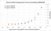
ARG57237 anti-5-hydroxymethylcytosine / 5-hmC antibody [RM236] ELISA image
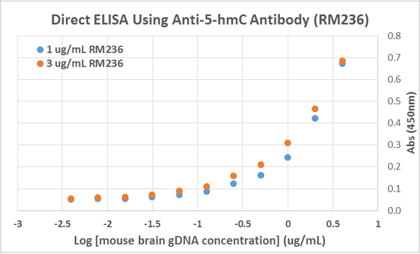
Direct ELISA: The plate was directly coated with different concentrations of genomic DNA isolated from Mouse brain tissue. 1 µg/ml or 3 µg/ml of ARG57237 anti-5-hydroxymethylcytosine / 5-hmC antibody [RM236] was used as the primary antibody, and a HRP-conjugated anti-rabbit IgG as the secondary antibody.
-

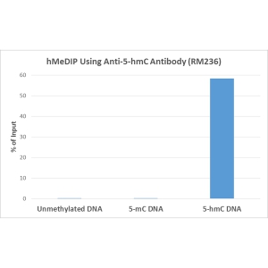
ARG57237 anti-5-hydroxymethylcytosine / 5-hmC antibody [RM236] hMeDIP image
hMeDIP: ARG57237 anti-5-hydroxymethylcytosine / 5-hmC antibody [RM236] at a 10:1 DNA:Ab ratio. 1 ng of unmethylated, 5-Methylcytosine (5-mC) or 5-Hydroxymethylcytosine (5-hmC) DNA standard (897 bp) was spiked in 1 µg of genomic DNA isolated from HeLa cells as the control. Realtime PCR was then performed to determine the capture of DNA standard as in % of input.