ARG55149
anti-AGAP4 antibody
anti-AGAP4 antibody for Flow cytometry,ICC/IF,IHC-Formalin-fixed paraffin-embedded sections,Western blot and Human
Signaling Transduction antibody
Overview
| Product Description | Rabbit Polyclonal antibody recognizes AGAP4 |
|---|---|
| Tested Reactivity | Hu |
| Tested Application | FACS, ICC/IF, IHC-P, WB |
| Host | Rabbit |
| Clonality | Polyclonal |
| Isotype | IgG |
| Target Name | AGAP4 |
| Antigen Species | Human |
| Immunogen | KLH-conjugated synthetic peptide corresponding to aa. 498-531 (C-terminus) of Human AGAP4. |
| Conjugation | Un-conjugated |
| Alternate Names | MRIP2; CTGLF5; AGAP-8; CTGLF1; AGAP-4; Arf-GAP with GTPase, ANK repeat and PH domain-containing protein 4; AGAP8; Centaurin-gamma-like family member 1; Centaurin-gamma-like family member 5 |
Application Instructions
| Application Suggestion |
|
||||||||||
|---|---|---|---|---|---|---|---|---|---|---|---|
| Application Note | * The dilutions indicate recommended starting dilutions and the optimal dilutions or concentrations should be determined by the scientist. | ||||||||||
| Positive Control | HT-1080 |
Properties
| Form | Liquid |
|---|---|
| Purification | Purification with Protein A and immunogen peptide. |
| Buffer | PBS and 0.09% (W/V) Sodium azide |
| Preservative | 0.09% (W/V) Sodium azide |
| Storage Instruction | For continuous use, store undiluted antibody at 2-8°C for up to a week. For long-term storage, aliquot and store at -20°C or below. Storage in frost free freezers is not recommended. Avoid repeated freeze/thaw cycles. Suggest spin the vial prior to opening. The antibody solution should be gently mixed before use. |
| Note | For laboratory research only, not for drug, diagnostic or other use. |
Bioinformation
| Database Links |
Swiss-port # Q96P64 Human Arf-GAP with GTPase, ANK repeat and PH domain-containing protein 4 |
|---|---|
| Gene Symbol | AGAP4 |
| Gene Full Name | ArfGAP with GTPase domain, ankyrin repeat and PH domain 4 |
| Function | Putative GTPase-activating protein. [UniProt] |
| Research Area | Signaling Transduction antibody |
| Calculated MW | 73 kDa |
Images (4) Click the Picture to Zoom In
-

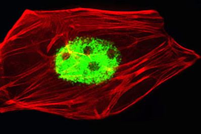
ARG55149 anti-AGAP4 antibody ICC/IF image
Immunofluorescence: HeLa cells stained with ARG55149 anti-AGAP4 antibody (green) at 1:25 dilution. Cytoplasmic actin was counterstained with Alexa Fluor® 555 conjugated with Phalloidin (red).
-
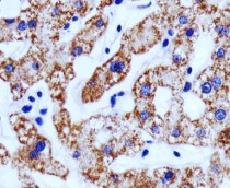
ARG55149 anti-AGAP4 antibody IHC-P image
Immunohistochemistry: Paraffin-embedded Human liver tissue stained with ARG55149 anti-AGAP4 antibody at 1:25 dilution.
-
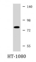
ARG55149 anti-AGAP4 antibody WB image
Western blot: 35 µg of HT-1080 cell lysate stained with ARG55149 anti-AGAP4 antibody at 1:1000 dilution.
-
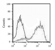
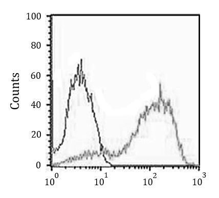
ARG55149 anti-AGAP4 antibody FACS image
Flow Cytometry: MCF7 cells stained with ARG55149 anti-AGAP4 antibody (right histogram) at 1:25 dilution or isotype control antibody (left histogram), followed by incubation with Alexa Fluor® 488 labelled secondary antibody.