ARG63338
anti-AIF1 / Iba 1 antibody
anti-AIF1 / Iba 1 antibody for ICC/IF,IHC-Formalin-fixed paraffin-embedded sections,Western blot and Bovine,Dog,Guinea pig,Human,Marmoset,Mouse,Rabbit,Rat
Cell Biology and Cellular Response antibody; Immune System antibody; Metabolism antibody; Neuroscience antibody; Activated Macrophage/Microglia Study antibody; Neuroinflammation Study antibody; Macroglial Marker antibody

Overview
| Product Description | Goat Polyclonal antibody recognizes AIF1 / Iba 1 |
|---|---|
| Tested Reactivity | Hu, Ms, Rat, Bov, Dog, Gpig, Marmoset, Rb |
| Predict Reactivity | Macaq, Pig |
| Tested Application | ICC/IF, IHC-P, WB |
| Specificity | This antibody is expected to recognize isoform 1 (NP_116573.1) and isoform 3 (NP_001614.3). AIF1 / IBA1 is thought to be involved in negative regulation of growth of vascular smooth muscle cells, which contributes to the anti-inflammatory response to vessel wall trauma. |
| Host | Goat |
| Clonality | Polyclonal |
| Isotype | IgG |
| Target Name | AIF1 / Iba 1 |
| Antigen Species | Human |
| Immunogen | C-TGPPAKKAISELP |
| Conjugation | Un-conjugated |
| Alternate Names | Ionized calcium-binding adapter molecule 1; Allograft inflammatory factor 1; IBA1; IRT-1; IRT1; AIF-1; Protein G1 |
Application Instructions
| Application Suggestion |
|
||||||||
|---|---|---|---|---|---|---|---|---|---|
| Application Note | WB: Recommend incubate at RT for 1h. IHC-P: Antigen Retrieval: Heat mediation was performed in Citrate buffer (pH 6.0). * The dilutions indicate recommended starting dilutions and the optimal dilutions or concentrations should be determined by the scientist. |
||||||||
| Positive Control | Raw264.7, Mouse brain and Rat brain | ||||||||
| Observed Size | 15 - 18 kDa |
Properties
| Form | Liquid |
|---|---|
| Purification | Purified from goat serum by antigen affinity chromatography. |
| Buffer | Tris saline (pH 7.3), 0.02% Sodium azide and 0.5% BSA. |
| Preservative | 0.02% Sodium azide |
| Stabilizer | 0.5% BSA |
| Concentration | 0.5 mg/ml |
| Storage Instruction | For continuous use, store undiluted antibody at 2-8°C for up to a week. For long-term storage, aliquot and store at -20°C or below. Storage in frost free freezers is not recommended. Avoid repeated freeze/thaw cycles. Suggest spin the vial prior to opening. The antibody solution should be gently mixed before use. |
| Note | For laboratory research only, not for drug, diagnostic or other use. |
Bioinformation
| Database Links | |
|---|---|
| Background | AIF1 / Iba 1 is a protein that binds actin and calcium. This gene is induced by cytokines and interferon and may promote macrophage activation and growth of vascular smooth muscle cells and T-lymphocytes. Polymorphisms in this gene may be associated with systemic sclerosis. Alternative splicing results in multiple transcript variants, but the full-length and coding nature of some of these variants is not certain. [provided by RefSeq, Jan 2016] |
| Function | AIF1 / Iba 1 is an Actin-binding protein. It enhances membrane ruffling and RAC activation. Enhances the actin-bundling activity of LCP1. Binds calcium. Plays a role in RAC signaling and in phagocytosis. May play a role in macrophage activation and function. Promotes the proliferation of vascular smooth muscle cells and of T-lymphocytes. Enhances lymphocyte migration. Plays a role in vascular inflammation. [UniProt] |
| Highlight | Related Antibody Duos and Panels: ARG30317 Activated Macrophage / Microglia Marker Antibody Duo Related products: AIF1 antibodies; AIF1 Duos / Panels; Anti-Goat IgG secondary antibodies; Related news: Microglial help TAM-ing inflammation in the brain LKB1 deficiency in T cells promotes gut tumors |
| Research Area | Cell Biology and Cellular Response antibody; Immune System antibody; Metabolism antibody; Neuroscience antibody; Activated Macrophage/Microglia Study antibody; Neuroinflammation Study antibody; Macroglial Marker antibody |
| Calculated MW | 17 kDa |
| PTM | Phosphorylated on serine residues. |
Images (4) Click the Picture to Zoom In
-
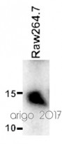
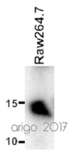
ARG63338 anti-AIF1 / Iba 1 antibody WB image
Western blot: 20 µg of Raw264.7 cell lysate stained with ARG63338 anti-AIF1 / Iba 1 antibody at 1:500 dilution.
-
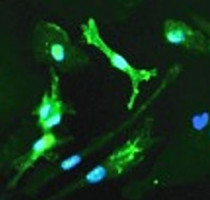
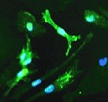
ARG63338 anti-AIF1 / Iba 1 antibody ICC/IF image
Immunofluorescence: LPS treated cultured Human microglia stained with ARG63338 anti-AIF1 / Iba 1 antibody at 1:100 dilution.
-
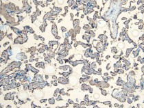
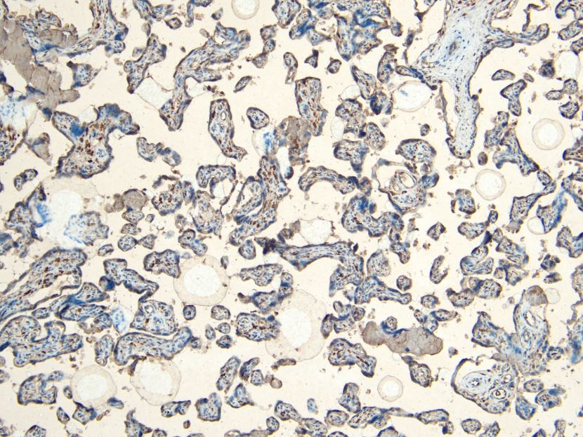
ARG63338 anti-AIF1 / Iba 1 antibody IHC-P image
Immunohistochemistry: Paraffin-embedded Human placenta tissue. Antigen Retrieval: Heat mediation was performed in Citrate buffer (pH 6.0). The tissue section was stained with ARG63338 anti-AIF1 / Iba 1 antibody at 6 µg/ml dilution followed by HRP-staining.
-
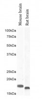
ARG63338 anti-AIF1 / Iba 1 antibody WB image
Western blot: 35 µg of Mouse brain and Rat brain lysates (in RIPA buffer) stained with ARG63338 anti-AIF1 / Iba 1 antibody at 1 µg/ml (left lane) or 2 µg/ml (right lane) and incubated at RT for 1 hour.

Customer's Feedback
Excellent

Review for anti-AIF1 / Iba 1 antibody
Application:WB
Sample:Raw264.7
Sample Loading Amount:20 µg
Primary Antibody Dilution Factor:1:500
Primary Antibody Incubation Time:overnight
Primary Antibody Incubation Temperature:4 ºC

Specific References
Sika Deer Velvet Antler Peptide Exerts Neuroprotective Effect in a Parkinson's Disease Model via Regulating Oxidative Damage and Gut Microbiota
IHC-P / Mouse


Porphyromonas gingivalis-induced periodontitis could contribute to cognitive impairment in Sprague–Dawley rats via the P38 MAPK signaling pathway
IHC-P / Rat
Effects of gingipain extracts on brain neuroinflammation in mice
IHC-P / Mouse
Activated STAT3 signaling pathway by ligature-induced periodontitis could contribute to neuroinflammation and cognitive impairment in rats
IHC-P / Rat
The imbalance of Th17/Treg via STAT3 activation modulates cognitive impairment in P. gingivalis LPS-induced periodontitis mice
IHC-Fr / Mouse
STAT3 signaling pathway could be involved in the progress of cognitive dysfunction caused by ligature-induced periodontitis.
IHC-P / Rat
Periodontitis Induced by P. gingivalis-LPS Is Associated With Neuroinflammation and Learning and Memory Impairment in Sprague-Dawley Rats
IHC-P / Rat
Effects of BIS-MEP on Reversing Amyloid Plaque Deposition and Spatial Learning and Memory Impairments in a Mouse Model of β-Amyloid Peptide- and Ibotenic Acid-Induced Alzheimer's Disease.
IHC-P / Mouse
Bis(9)-(-)-Meptazinol, a novel dual-binding AChE inhibitor, rescues cognitive deficits and pathological changes in APP/PS1 transgenic mice.
IHC-P / Mouse
Porphyromonas gingivalis lipopolysaccharide induces cognitive dysfunction, mediated by neuronal inflammation via activation of the TLR4 signaling pathway in C57BL/6 mice.
IHC-P / Mouse


