ARG58219
anti-AMFR antibody
anti-AMFR antibody for Flow cytometry,IHC-Formalin-fixed paraffin-embedded sections,Western blot and Human,Mouse,Rat
Overview
| Product Description | Rabbit Polyclonal antibody recognizes AMFR |
|---|---|
| Tested Reactivity | Hu, Ms, Rat |
| Tested Application | FACS, IHC-P, WB |
| Host | Rabbit |
| Clonality | Polyclonal |
| Isotype | IgG |
| Target Name | AMFR |
| Antigen Species | Human |
| Immunogen | E. coli-derived Human AMFR recombinant protein (Position: E553-S643). Human AMFR shares 89% amino acid (aa) sequence identity with Mouse AMFR. |
| Conjugation | Un-conjugated |
| Alternate Names | EC 6.3.2.-; E3 ubiquitin-protein ligase AMFR; RNF45; GP78; Autocrine motility factor receptor; gp78; AMF receptor; RING finger protein 45 |
Application Instructions
| Application Suggestion |
|
||||||||
|---|---|---|---|---|---|---|---|---|---|
| Application Note | IHC-P: Antigen Retrieval: Heat mediation was performed in Citrate buffer (pH 6.0) for 20 min. * The dilutions indicate recommended starting dilutions and the optimal dilutions or concentrations should be determined by the scientist. |
Properties
| Form | Liquid |
|---|---|
| Purification | Affinity purification with immunogen. |
| Buffer | 0.9% NaCl, 0.2% Na2HPO4, 0.05% Sodium azide and 5% BSA. |
| Preservative | 0.05% Sodium azide |
| Stabilizer | 5% BSA |
| Concentration | 0.5 mg/ml |
| Storage Instruction | For continuous use, store undiluted antibody at 2-8°C for up to a week. For long-term storage, aliquot and store at -20°C or below. Storage in frost free freezers is not recommended. Avoid repeated freeze/thaw cycles. Suggest spin the vial prior to opening. The antibody solution should be gently mixed before use. |
| Note | For laboratory research only, not for drug, diagnostic or other use. |
Bioinformation
| Database Links | |
|---|---|
| Gene Symbol | AMFR |
| Gene Full Name | autocrine motility factor receptor, E3 ubiquitin protein ligase |
| Background | This locus encodes a glycosylated transmembrane receptor. Its ligand, autocrine motility factor, is a tumor motility-stimulating protein secreted by tumor cells. The encoded receptor is also a member of the E3 ubiquitin ligase family of proteins. It catalyzes ubiquitination and endoplasmic reticulum-associated degradation of specific proteins. [provided by RefSeq, Feb 2012] |
| Function | E3 ubiquitin-protein ligase that mediates the polyubiquitination of a number of proteins such as CD3D, CYP3A4, CFTR and APOB for proteasomal degradation. Component of a VCP/p97-AMFR/gp78 complex that participates in the final step of endoplasmic reticulum-associated degradation (ERAD). The VCP/p97-AMFR/gp78 complex is involved in the sterol-accelerated ERAD degradation of HMGCR through binding to the HMGCR-INSIG complex at the ER membrane and initiating ubiquitination of HMGCR. The ubiquitinated HMGCR is then released from the ER by the complex into the cytosol for subsequent destruction. Also acts as a scaffold protein to assemble a complex that couples ubiquitination, retranslocation and deglycosylation. Mediates tumor invasion and metastasis as a receptor for the GPI/autocrine motility factor. [UniProt] |
| Cellular Localization | Endoplasmic reticulum membrane; Multi-pass membrane protein. [UniProt] |
| Calculated MW | 73 kDa |
Images (6) Click the Picture to Zoom In
-
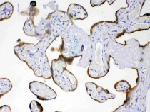
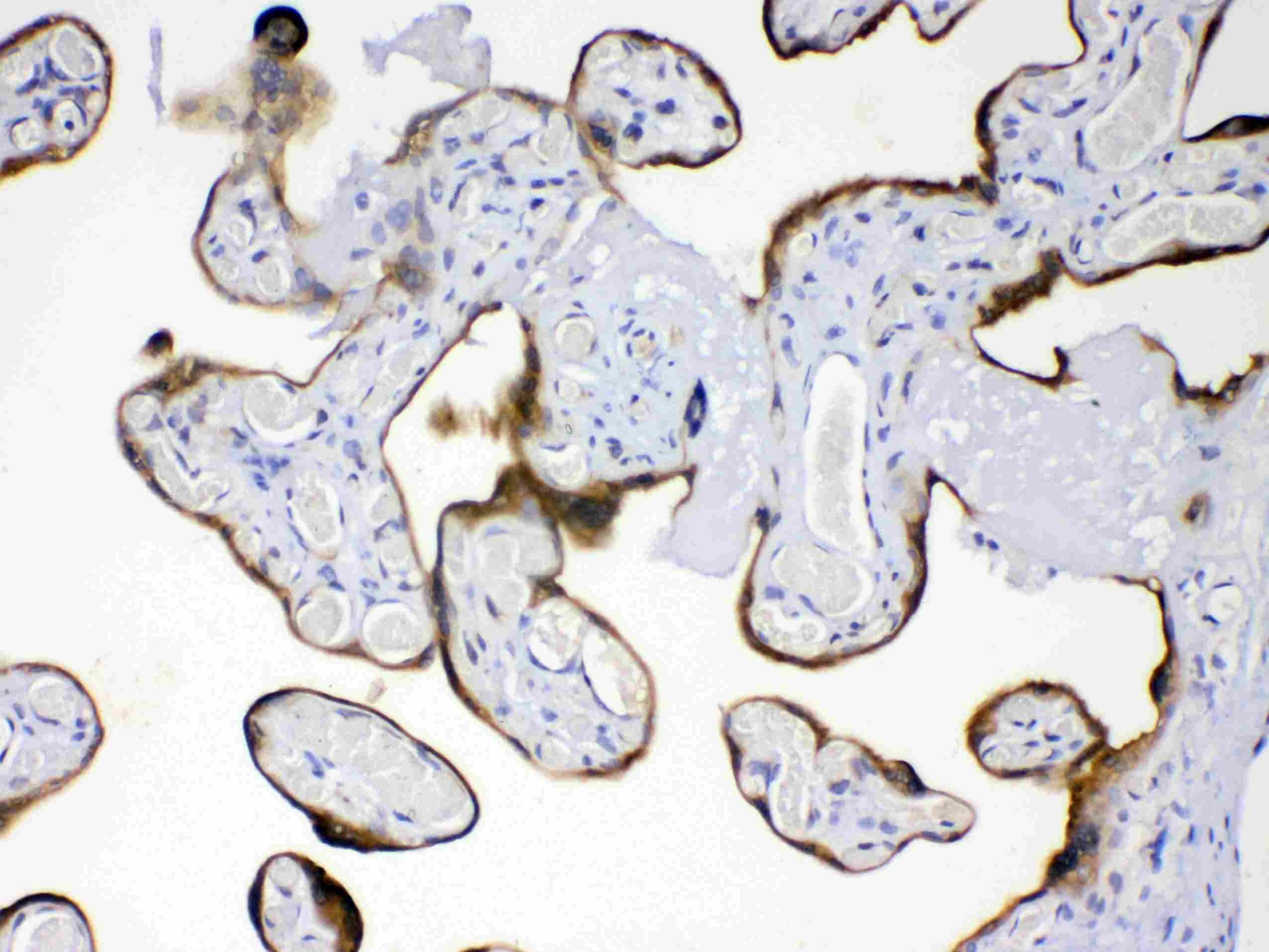
ARG58219 anti-AMFR antibody IHC-P image
Immunohistochemistry: Paraffin-embedded Human placentas stained with ARG58219 anti-AMFR antibody at 1 µg/ml dilution.
-
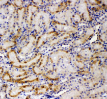
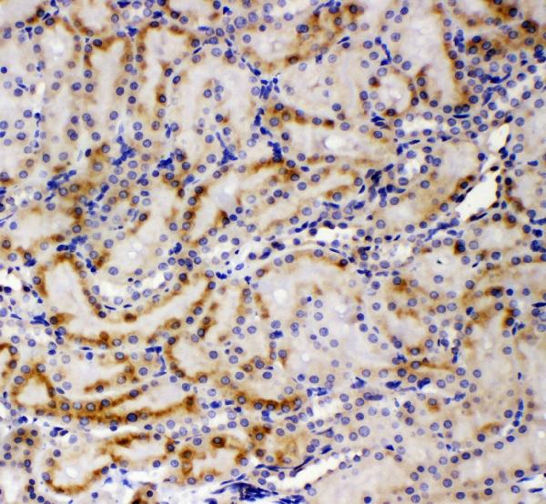
ARG58219 anti-AMFR antibody IHC-P image
Immunohistochemistry: Paraffin-embedded Rat kidney tissue. Antigen Retrieval: Heat mediation was performed in Citrate buffer (pH 6.0) for 20 min. The tissue section was blocked with 10% goat serum. The tissue section was then stained with ARG58219 anti-AMFR antibody at 1 µg/ml dilution, overnight at 4°C.
-
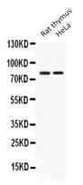
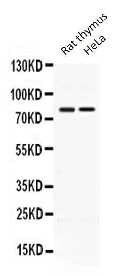
ARG58219 anti-AMFR antibody WB image
Western blot: Rat thymus extract and HeLa whole cell lysate stained with ARG58219 anti-AMFR antibody at 0.5 µg/ml dilution.
-
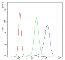
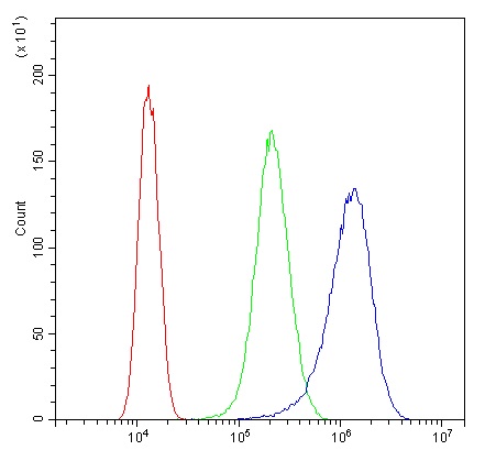
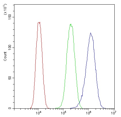
ARG58219 anti-AMFR antibody FACS image
Flow Cytometry: SiHa cells were blocked with 10% normal goat serum and then stained with ARG58219 anti-AMFR antibody (blue) at 1 µg/10^6 cells for 30 min at 20°C, followed by incubation with DyLight®488 labelled secondary antibody. Isotype control antibody (green) was rabbit IgG (1 µg/10^6 cells) used under the same conditions. Unlabelled sample (red) was also used as a control.
-
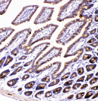
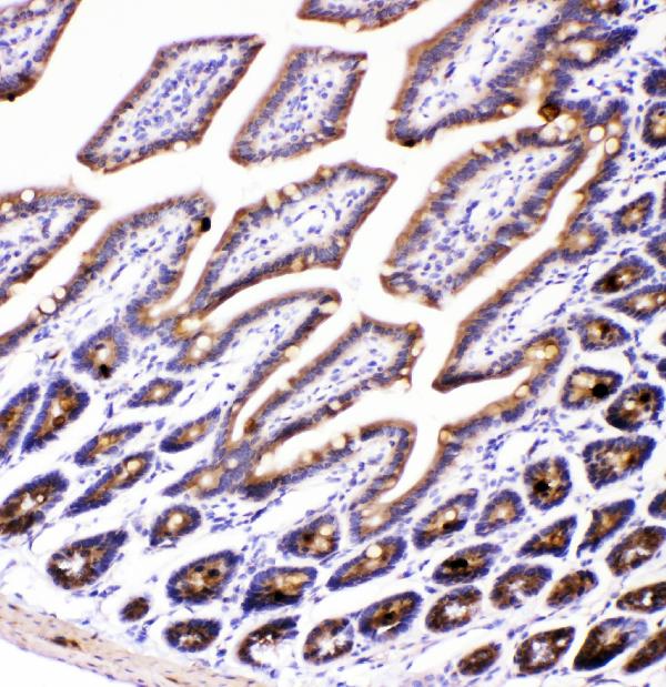
ARG58219 anti-AMFR antibody IHC-P image
Immunohistochemistry: Paraffin-embedded Mouse intestine tissue. Antigen Retrieval: Heat mediation was performed in Citrate buffer (pH 6.0) for 20 min. The tissue section was blocked with 10% goat serum. The tissue section was then stained with ARG58219 anti-AMFR antibody at 1 µg/ml dilution, overnight at 4°C.
-
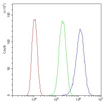
ARG58219 anti-AMFR antibody FACS image
Flow Cytometry: U87 cells were blocked with 10% normal goat serum and then stained with ARG58219 anti-AMFR antibody (blue) at 1 µg/10^6 cells for 30 min at 20°C, followed by incubation with DyLight®488 labelled secondary antibody. Isotype control antibody (green) was rabbit IgG (1 µg/10^6 cells) used under the same conditions. Unlabelled sample (red) was also used as a control.