ARG63742
anti-ASPN / Asporin antibody
anti-ASPN / Asporin antibody for ICC/IF,Western blot and Human,Mouse,Rat
Signaling Transduction antibody
Overview
| Product Description | Goat Polyclonal antibody recognizes ASPN / Asporin |
|---|---|
| Tested Reactivity | Hu, Ms, Rat |
| Tested Application | ICC/IF, WB |
| Host | Goat |
| Clonality | Polyclonal |
| Isotype | IgG |
| Target Name | ASPN / Asporin |
| Antigen Species | Human |
| Immunogen | C-IHENKVKKIQKDT |
| Conjugation | Un-conjugated |
| Alternate Names | Periodontal ligament-associated protein 1; OS3; SLRR1C; Asporin; PLAP-1; PLAP1 |
Application Instructions
| Application Suggestion |
|
||||||
|---|---|---|---|---|---|---|---|
| Application Note | WB: Recommend incubate at RT for 1h. * The dilutions indicate recommended starting dilutions and the optimal dilutions or concentrations should be determined by the scientist. |
Properties
| Form | Liquid |
|---|---|
| Purification | Purified from goat serum by antigen affinity chromatography. |
| Buffer | Tris saline (pH 7.3), 0.02% Sodium azide and 0.5% BSA. |
| Preservative | 0.02% Sodium azide |
| Stabilizer | 0.5% BSA |
| Concentration | 0.5 mg/ml |
| Storage Instruction | For continuous use, store undiluted antibody at 2-8°C for up to a week. For long-term storage, aliquot and store at -20°C or below. Storage in frost free freezers is not recommended. Avoid repeated freeze/thaw cycles. Suggest spin the vial prior to opening. The antibody solution should be gently mixed before use. |
| Note | For laboratory research only, not for drug, diagnostic or other use. |
Bioinformation
| Database Links | |
|---|---|
| Background | This gene encodes a cartilage extracellular protein that is member of the small leucine-rich proteoglycan family. The encoded protein may regulate chondrogenesis by inhibiting transforming growth factor-beta 1-induced gene expression in cartilage. This protein also binds collagen and calcium and may induce collagen mineralization. Polymorphisms in the aspartic acid repeat region of this gene are associated with a susceptibility to osteoarthritis. Alternate splicing results in multiple transcript variants.[provided by RefSeq, Jul 2010] |
| Research Area | Signaling Transduction antibody |
| Calculated MW | 43 kDa |
| PTM | There is no serine/glycine dipeptide sequence expected for the attachment of O-linked glycosaminoglycans and this is probably not a proteoglycan. The O-linked polysaccharide on 54-Ser is probably the mucin type linked to GalNAc. The N-linked glycan at Asn-282 is composed of variable structures of GlcNAc, mannose, fucose, HexNAc and hexose. |
Images (3) Click the Picture to Zoom In
-
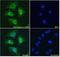
ARG63742 anti-ASPN / Asporin antibody ICC/IF image
Immunofluorescence: Paraformaldehyde fixed HeLa cells permeabilized with 0.15% Triton. Cells were stained with ARG63742 anti-ASPN / Asporin antibody (green) at 10 µg/ml dilution for 1 hour. DAPI (blue) for nuclear staining. Negative control: Unimmunized goat IgG (green) at 10 µg/ml dilution.
-
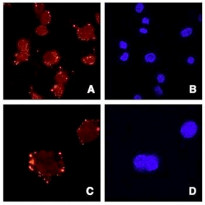
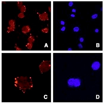
ARG63742 anti-ASPN / Asporin antibody ICC/IF image
Immunofluorescence: ATDC5 cells (Panels A and C) and DAPI (panels B and D) stained with ARG63742 anti-ASPN / Asporin antibody. Data gratefuly received from Dr. Shiro Ikegawa, SNP Research Center, RIKEN, Japan.
-
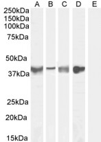
ARG63742 anti-ASPN / Asporin antibody WB image
Western blot: 35 µg of Human tonsil (A), Human uterus (B), Mouse skeletal muscle (C), Rat skeletal muscle (D) and Human cerebellum (E, negative control) lysates (in RIPA buffer) stained with ARG63742 anti-ASPN / Asporin antibody at 0.1 µg/ml (A), 0.3 µg/ml (B, C) and 1 µg/ml (D, E) dilutions and incubated at RT for 1 hour.