ARG43715
anti-CD4 antibody
anti-CD4 antibody for Flow cytometry,IHC-Formalin-fixed paraffin-embedded sections,Western blot and Mouse
Overview
| Product Description | Rabbit Polyclonal antibody recognizes CD4 |
|---|---|
| Tested Reactivity | Ms |
| Predict Reactivity | Rat |
| Tested Application | FACS, IHC-P, WB |
| Host | Rabbit |
| Clonality | Polyclonal |
| Isotype | IgG |
| Target Name | CD4 |
| Antigen Species | Rat |
| Immunogen | Recombinant protein corresponding to K28-I457 of Rat CD4. |
| Conjugation | Un-conjugated |
| Protein Full Name | T-cell surface glycoprotein CD4 |
| Alternate Names | CD4mut; CD antigen CD4; T-cell surface glycoprotein CD4; T-cell surface antigen T4/Leu-3; p55; W3/25 |
Application Instructions
| Application Suggestion |
|
||||||||
|---|---|---|---|---|---|---|---|---|---|
| Application Note | IHC-P: Antigen Retrieval: By heat mediation. * The dilutions indicate recommended starting dilutions and the optimal dilutions or concentrations should be determined by the scientist. |
||||||||
| Positive Control | mouse PBMC, Raw264.7, ANA-1, mouse thymus tissue | ||||||||
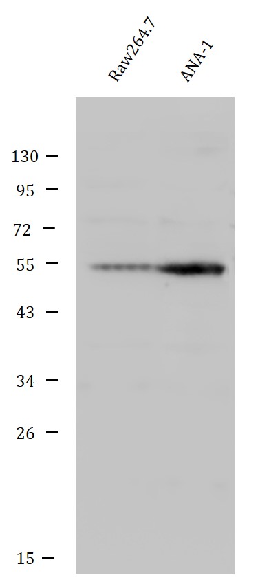
| Observed Size | 54 kDa |
Properties
| Form | Liquid |
|---|---|
| Purification | Affinity purification with immunogen. |
| Buffer | 0.2% Na2HPO4, 0.9% NaCl, 0.01% Sodium azide and 4% Trehalose. |
| Preservative | 0.01% Sodium azide |
| Stabilizer | 4% Trehalose |
| Concentration | 0.5 mg/ml |
| Storage Instruction | For continuous use, store undiluted antibody at 2-8°C for up to a week. For long-term storage, aliquot and store at -20°C or below. Storage in frost free freezers is not recommended. Avoid repeated freeze/thaw cycles. Suggest spin the vial prior to opening. The antibody solution should be gently mixed before use. |
| Note | For laboratory research only, not for drug, diagnostic or other use. |
Bioinformation
| Database Links | |
|---|---|
| Gene Symbol | Cd4 |
| Gene Full Name | Cd4 molecule |
| Background | This gene encodes a membrane glycoprotein of T lymphocytes that interacts with major histocompatibility complex class II antigenes and is also a receptor for the human immunodeficiency virus. This gene is expressed not only in T lymphocytes, but also in B cells, macrophages, and granulocytes. It is also expressed in specific regions of the brain. The protein functions to initiate or augment the early phase of T-cell activation, and may function as an important mediator of indirect neuronal damage in infectious and immune-mediated diseases of the central nervous system. Multiple alternatively spliced transcript variants encoding different isoforms have been identified in this gene. [provided by RefSeq, Aug 2010] |
| Function | Integral membrane glycoprotein that plays an essential role in the immune response and serves multiple functions in responses against both external and internal offenses. In T-cells, functions primarily as a coreceptor for MHC class II molecule:peptide complex. The antigens presented by class II peptides are derived from extracellular proteins while class I peptides are derived from cytosolic proteins. Interacts simultaneously with the T-cell receptor (TCR) and the MHC class II presented by antigen presenting cells (APCs). In turn, recruits the Src kinase LCK to the vicinity of the TCR-CD3 complex. LCK then initiates different intracellular signaling pathways by phosphorylating various substrates ultimately leading to lymphokine production, motility, adhesion and activation of T-helper cells. In other cells such as macrophages or NK cells, plays a role in differentiation/activation, cytokine expression and cell migration in a TCR/LCK-independent pathway. Participates in the development of T-helper cells in the thymus and triggers the differentiation of monocytes into functional mature macrophages. (Microbial infection) Primary receptor for human immunodeficiency virus-1 (HIV-1) (PubMed:2214026, PubMed:16331979, PubMed:9641677, PubMed:12089508). Down-regulated by HIV-1 Vpu (PubMed:17346169). Acts as a receptor for Human Herpes virus 7/HHV-7 (PubMed:7909607). [UniProt] |
| Cellular Localization | Cell membrane; Single-pass type I membrane protein. Note=Localizes to lipid rafts (PubMed:12517957, PubMed:9168119). Removed from plasma membrane by HIV-1 Nef protein that increases clathrin-dependent endocytosis of this antigen to target it to lysosomal degradation. Cell surface expression is also down-modulated by HIV-1 Envelope polyprotein gp160 that interacts with, and sequesters CD4 in the endoplasmic reticulum. [UniProt] |
| Calculated MW | 51 kDa |
| PTM | Palmitoylation and association with LCK contribute to the enrichment of CD4 in lipid rafts. [UniProt] |
Images (3) Click the Picture to Zoom In
-
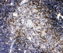
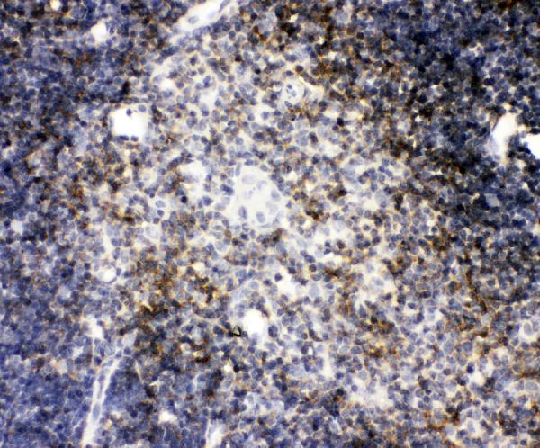
ARG43715 anti-CD4 antibody IHC-P image
Immunohistochemistry: Paraffin-embedded Mouse thymus tissue. Antigen Retrieval: Heat mediation was performed in EDTA buffer (pH 8.0). The tissue section was blocked with 10% goat serum. The tissue section was then stained with ARG43715 anti-CD4 antibody at 1 µg/ml dilution, and incubated overnight at 4°C.
-
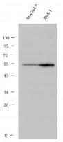
ARG43715 anti-CD4 antibody WB image
Western blot: 50 µg of samples under reducing conditions. Mouse Raw264.7 and ANA-1 whole cell lysates stained with ARG43715 anti-CD4 antibody at 0.5 µg/ml dilution, and incubated overnight at 4°C.
-
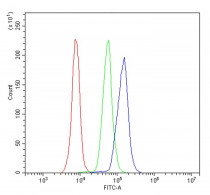
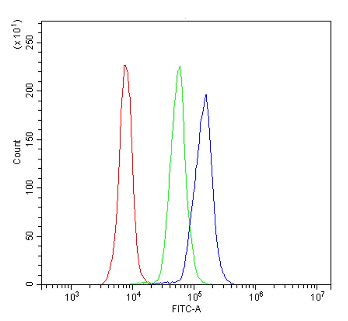
ARG43715 anti-CD4 antibody FACS image
Flow Cytometry: Mouse PBMC cells were blocked with 10% normal goat serum and then stained with ARG43715 anti-CD4 antibody (blue) at 1 µg/10^6 cells for 30 min at 20°C, followed by incubation with DyLight®488 labelled secondary antibody. Isotype control antibody (green) was rabbit IgG (1 µg/10^6 cells) used under the same conditions. Unlabelled sample (red) was also used as a control.