ARG54337
anti-CIDE A antibody
anti-CIDE A antibody for IHC-Formalin-fixed paraffin-embedded sections,Western blot and Human,Mouse
Cell Biology and Cellular Response antibody; Cell Death antibody; Gene Regulation antibody; Metabolism antibody
Overview
| Product Description | Rabbit Polyclonal antibody recognizes CIDE A |
|---|---|
| Tested Reactivity | Hu, Ms |
| Tested Application | IHC-P, WB |
| Specificity | This antibody recognizes mouse CIDE-A (25kDa) and does not cross-react with CIDE-B. |
| Host | Rabbit |
| Clonality | Polyclonal |
| Isotype | IgG |
| Target Name | CIDE A |
| Antigen Species | Mouse |
| Immunogen | Peptide corresponding to aa 200-214 of mouse CIDE-A (accession no. AAC34985). |
| Conjugation | Un-conjugated |
| Alternate Names | CIDE-A; Cell death-inducing DFFA-like effector A; Cell death activator CIDE-A |
Application Instructions
| Application Suggestion |
|
||||||
|---|---|---|---|---|---|---|---|
| Application Note | * The dilutions indicate recommended starting dilutions and the optimal dilutions or concentrations should be determined by the scientist. | ||||||
| Positive Control | Mouse heart |
Properties
| Form | Liquid |
|---|---|
| Purification | Immunoaffinity chroma-tography |
| Buffer | PBS (pH 7.4) and 0.02% Sodium azide |
| Preservative | 0.02% Sodium azide |
| Storage Instruction | For continuous use, store undiluted antibody at 2-8°C for up to a week. For long-term storage, aliquot and store at -20°C or below. Storage in frost free freezers is not recommended. Avoid repeated freeze/thaw cycles. Suggest spin the vial prior to opening. The antibody solution should be gently mixed before use. |
| Note | For laboratory research only, not for drug, diagnostic or other use. |
Bioinformation
| Database Links | |
|---|---|
| Gene Symbol | Cidea |
| Gene Full Name | cell death-inducing DNA fragmentation factor, alpha subunit-like effector A |
| Background | DFF45-related proteins CIDE-A and CIDE-B have recently been identified. CIDE contains a new type of domain termed CIDE-N, which has high homology with the regulatory domains of DFF45/ICAD and DFF40/CAD. Expression of CIDE-A induces DNA fragmentation and activates apoptosis, which is inhibited by DFF45. CIDE-A is a DFF45-inhibitable effector that promotes cell death and DNA fragmentation. CIDE-A is expressed in many tissues. |
| Function | Binds to lipid droplets and regulates their enlargement, thereby restricting lipolysis and favoring storage. At focal contact sites between lipid droplets, promotes directional net neutral lipid transfer from the smaller to larger lipid droplets. The transfer direction may be driven by the internal pressure difference between the contacting lipid droplet pair and occurs at a lower rate than that promoted by CIDEC. Acts as a CEBPB coactivator in mammary epithelial cells to control the expression of a subset of CEBPB downstream target genes, including ID2, IGF1, PRLR, SOCS1, SOCS3, XDH, but not casein. By interacting with CEBPB, strengthens the association of CEBPB with the XDH promoter, increases histone acetylation and dissociates HDAC1 from the promoter. When overexpressed, induces apoptosis. The physiological significance of its role in apoptosis is unclear. [UniProt] |
| Research Area | Cell Biology and Cellular Response antibody; Cell Death antibody; Gene Regulation antibody; Metabolism antibody |
| Calculated MW | 25 kDa |
Images (3) Click the Picture to Zoom In
-
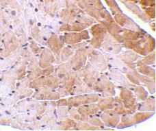
ARG54337 anti-CIDE A antibody IHC-P image
Immunohistochemistry: Human heart tissue stained with ARG54337 anti-CIDE A antibody at 2.5 μg/ml dilution.
-
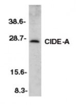
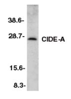
ARG54337 anti-CIDE A antibody WB image
Western blot: mouse heart tissue stained with ARG54337 anti-CIDE A antibody at 2 μg/ml dilution.
-

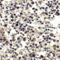
ARG54337 anti-CIDE A antibody IHC-P image
Immunohistochemistry: Mouse heart tissue stained with ARG54337 anti-CIDE A antibody at 5 μg/ml dilution.