ARG10703
anti-Calretinin antibody [6A9]
anti-Calretinin antibody [6A9] for ICC/IF,IHC-Frozen sections,Western blot and Human,Mouse,Rat,Cow

Overview
| Product Description | Mouse Monoclonal antibody [6A9] recognizes Calretinin |
|---|---|
| Tested Reactivity | Hu, Ms, Rat, Cow |
| Tested Application | ICC/IF, IHC-Fr, WB |
| Host | Mouse |
| Clonality | Monoclonal |
| Clone | 6A9 |
| Isotype | IgA |
| Target Name | Calretinin |
| Antigen Species | Human |
| Immunogen | Full-length recombinant Human protein. |
| Conjugation | Un-conjugated |
| Alternate Names | CAB29; CR; CAL2; 29 kDa calbindin; Calretinin |
Application Instructions
| Application Suggestion |
|
||||||||
|---|---|---|---|---|---|---|---|---|---|
| Application Note | * The dilutions indicate recommended starting dilutions and the optimal dilutions or concentrations should be determined by the scientist. |
Properties
| Form | Liquid |
|---|---|
| Purification | Affinity purification. |
| Buffer | PBS, 5 mM Sodium azide and 50% Glycerol. |
| Preservative | 5 mM Sodium azide |
| Stabilizer | 50% Glycerol |
| Concentration | 1 mg/ml |
| Storage Instruction | For continuous use, store undiluted antibody at 2-8°C for up to a week. For long-term storage, aliquot and store at -20°C. Storage in frost free freezers is not recommended. Avoid repeated freeze/thaw cycles. Suggest spin the vial prior to opening. The antibody solution should be gently mixed before use. |
| Note | For laboratory research only, not for drug, diagnostic or other use. |
Bioinformation
| Database Links | |
|---|---|
| Gene Symbol | CALB2 |
| Gene Full Name | calbindin 2 |
| Background | This gene encodes an intracellular calcium-binding protein belonging to the troponin C superfamily. Members of this protein family have six EF-hand domains which bind calcium. This protein plays a role in diverse cellular functions, including message targeting and intracellular calcium buffering. It also functions as a modulator of neuronal excitability, and is a diagnostic marker for some human diseases, including Hirschsprung disease and some cancers. Alternative splicing results in multiple transcript variants. [provided by RefSeq, Jun 2010] |
| Function | Calretinin is a calcium-binding protein which is abundant in auditory neurons. [UniProt] |
| Calculated MW | 32 kDa |
Images (7) Click the Picture to Zoom In
-
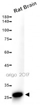
ARG10703 anti-Calretinin antibody [6A9] WB image
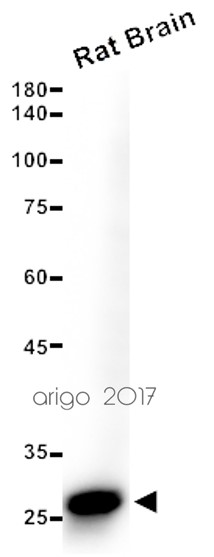
Western blot: 15 µg of Rat brain lysate stained with ARG10703 anti-Calretinin antibody [6A9] at 1:2500 dilution.
-
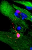
ARG10703 anti-Calretinin antibody [6A9] ICC/IF image
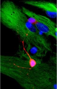
Immunocytochemistry: Rat mixed neuron / glial cultures stained with ARG10703 anti-Calretinin antibody [6A9] (red) at 1:2000, and co-stained with chicken polyclonal antibody to Vimentin (green). Calretinin is prominently expressed in small number of interneurons, while astrocytes and fibroblasts were visualized with the vimentin antibody.
-
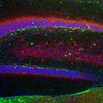
ARG10703 anti-Calretinin antibody [6A9] IHC-Fr image
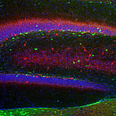
Immunohistochemistry: Frozen section of Rat hippocampus stained with ARG10703 anti-Calretinin antibody [6A9] (green) at 1:2000 dilution and costained with ARG10686 anti-Parvalbumin antibody (red) at 1:1000 dilution. DAPI (blue) for nuclear staining. (Sample preparation: Following transcardial perfusion of Rat with 4% paraformaldehyde, brain was post fixed for 24 hours, cut to 45 µM, and free-floating sections were stained with above antibodies.)
-
ARG10703 anti-Calretinin antibody [6A9] IHC-Fr image
Immunohistochemistry: Frozen sections of adult Rat cortical (45 µM; fixed by transcardial perfusion with 4% paraformaldehyde) was stained with ARG10703 anti-Calretinin antibody [6A9] at 1:1000 (red) and co-stained with chicken polyclonal antibody to calbinidin (green). In the motor cortex, calretinin is expressed in a small population of interneurons that do not express calbindin. Because each antibody specifically labels a different population of cells exclusively, the cells are either stained with red, or green.
-
ARG10703 anti-Calretinin antibody [6A9] WB image
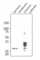
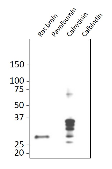
Western blot: Rat brain, pavalbumin, calretinin, and calbindin recombinant proteins was stained with ARG10703 anti-Calretinin antibody [6A9] at 1:5000 dilution.
-
ARG10703 anti-Calretinin antibody [6A9] WB image
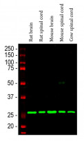
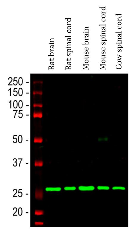
Western blot: Rat brain, Rat spinal cord, Mouse brain, Mouse spinal cord and Cow spinal cord lysates stained with ARG10703 anti-Calretinin antibody [6A9] (green) at 1:2000 dilution.
-

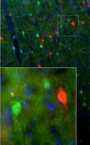
ARG10703 anti-Calretinin antibody [6A9] IHC-Fr image
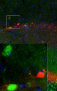
Immunohistochemistry: Frozen sections of adult Mouse brain hippocampal (45 µM; fixed by transcardial perfusion with 4% paraformaldehyde) was stained with ARG10703 anti-Calretinin antibody [6A9] at 1:1000 (red) and co-stained with antibody to calbinidin (green). In the stratum radiatum of CA1 region, calretinin expresses in a small number of interneurons that do not express calbindin. As a result, our antibodies label different neurons in either red or green. Insets are high-magnification images of the boxed area in each picture. Blue is a hoechst DNA staining.

Customer's Feedback
Excellent

Review for anti-Calretinin antibody [6A9]
Application:WB
Sample:Rat brain
Sample Loading Amount:15 µg
Primary Antibody Dilution Factor:1:2500
Primary Antibody Incubation Time:overnight
Primary Antibody Incubation Temperature:4 ºC

Excellent

Review for anti-Calretinin antibody [6A9]
Application:WB
Sample:Mouse Cerebellum
Sample Loading Amount:15 µg
Primary Antibody Dilution Factor:1:2500
Primary Antibody Incubation Time:overnight
Primary Antibody Incubation Temperature:4 ºC


