ARG41725
anti-Caspase 4 antibody
anti-Caspase 4 antibody for Flow cytometry,ICC/IF,IHC-Formalin-fixed paraffin-embedded sections,Western blot and Human,Mouse,Rat
Overview
| Product Description | Rabbit Polyclonal antibody recognizes Caspase 4 |
|---|---|
| Tested Reactivity | Hu, Ms, Rat |
| Tested Application | FACS, ICC/IF, IHC-P, WB |
| Host | Rabbit |
| Clonality | Polyclonal |
| Isotype | IgG |
| Target Name | Caspase 4 |
| Antigen Species | Mouse |
| Immunogen | Recombinant protein corresponding to E108-T195 of Mouse Caspase 4. |
| Conjugation | Un-conjugated |
| Alternate Names | Mih1/TX; Protease ICH-2; Caspase-4; EC 3.4.22.57; TX; CASP-4; Protease TX; ICE; ICH-2; ICE(rel)II; rel; ICEREL-II |
Application Instructions
| Application Suggestion |
|
||||||||||
|---|---|---|---|---|---|---|---|---|---|---|---|
| Application Note | IHC-P: Antigen Retrieval: Heat mediation was performed in Citrate buffer (pH 6.0, epitope retrieval solution) for 20 min. * The dilutions indicate recommended starting dilutions and the optimal dilutions or concentrations should be determined by the scientist. |
||||||||||
| Observed Size | ~ 43 kDa |
Properties
| Form | Liquid |
|---|---|
| Purification | Affinity purification with immunogen. |
| Buffer | 0.2% Na2HPO4, 0.9% NaCl, 0.05% Sodium azide and 4% Trehalose. |
| Preservative | 0.05% Sodium azide |
| Stabilizer | 4% Trehalose |
| Concentration | 0.5 mg/ml |
| Storage Instruction | For continuous use, store undiluted antibody at 2-8°C for up to a week. For long-term storage, aliquot and store at -20°C or below. Storage in frost free freezers is not recommended. Avoid repeated freeze/thaw cycles. Suggest spin the vial prior to opening. The antibody solution should be gently mixed before use. |
| Note | For laboratory research only, not for drug, diagnostic or other use. |
Bioinformation
| Database Links | |
|---|---|
| Gene Symbol | CASP4 |
| Gene Full Name | caspase 4, apoptosis-related cysteine peptidase |
| Background | This gene encodes a protein that is a member of the cysteine-aspartic acid protease (caspase) family. Sequential activation of caspases plays a central role in the execution-phase of cell apoptosis. Caspases exist as inactive proenzymes composed of a prodomain and a large and small protease subunit. Activation of caspases requires proteolytic processing at conserved internal aspartic residues to generate a heterodimeric enzyme consisting of the large and small subunits. This caspase is able to cleave and activate its own precursor protein, as well as caspase 1 precursor. When overexpressed, this gene induces cell apoptosis. Alternative splicing results in transcript variants encoding distinct isoforms. [provided by RefSeq, Jul 2008] |
| Function | Involved in the activation cascade of caspases responsible for apoptosis execution. Involved in ER-stress induced apoptosis. Cleaves caspase-1. [UniProt] |
| Cellular Localization | Cytoplasm, cytosol. Endoplasmic reticulum membrane; Peripheral membrane protein; Cytoplasmic side. Mitochondrion. Inflammasome. Secreted. Note=Predominantly localizes to the endoplasmic reticulum (ER). Association with the ER membrane requires TMEM214 (PubMed:15123740). Released in the extracellular milieu by keratinocytes following UVB irradiation (PubMed:22246630). [UniProt] |
| Calculated MW | 43 kDa |
| PTM | In response to activation signals, including endoplasmic reticulum stress or treatment with amyloid beta A4 protein fragments (such as beta-amyloid protein 40), undergoes autoproteolytic cleavage. [UniProt] |
Images (5) Click the Picture to Zoom In
-

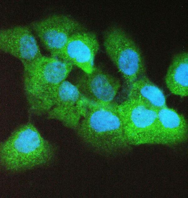
ARG41725 anti-Caspase 4 antibody ICC/IF image
Immunofluorescence: A431 cells stained with ARG41725 anti-Caspase 4 antibody at 2 µg/ml dilution, overnight at 4°C. DAPI (blue) for nuclear staining.
-

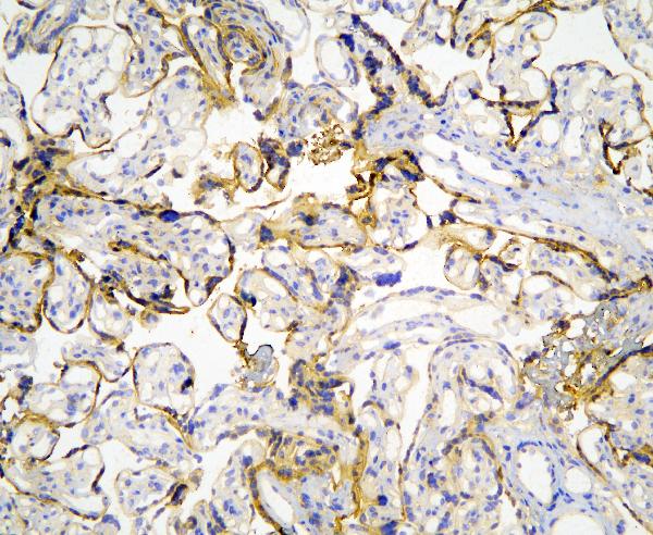
ARG41725 anti-Caspase 4 antibody IHC-P image
Immunohistochemistry: Paraffin-embedded Human placenta tissue. Antigen Retrieval: Heat mediation was performed in Citrate buffer (pH 6.0, epitope retrieval solution) for 20 min. The tissue section was blocked with 10% goat serum. The tissue section was then stained with ARG41725 anti-Caspase 4 antibody at 1 µg/ml dilution, overnight at 4°C.
-
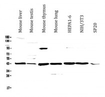
ARG41725 anti-Caspase 4 antibody WB image
Western blot: 50 µg of samples under reducing conditions. Mouse liver, Mouse testis, Mouse thymus, Mouse lung, HEPA1-6, NIH/3T3 and SP20 whole cell lysates stained with ARG41725 anti-Caspase 4 antibody at 0.5 µg/ml dilution, overnight at 4°C.
-
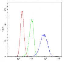
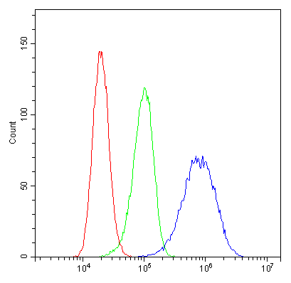
ARG41725 anti-Caspase 4 antibody FACS image
Flow Cytometry: A431 cells were blocked with 10% normal goat serum and then stained with ARG41725 anti-Caspase 4 antibody (blue) at 1 µg/10^6 cells for 30 min at 20°C, followed by incubation with DyLight®488 labelled secondary antibody. Isotype control antibody (green) was Rabbit IgG (1 µg/10^6 cells) used under the same conditions. Unlabelled sample (red) was also used as a control.
-
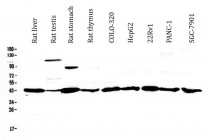
ARG41725 anti-Caspase 4 antibody WB image
Western blot: 50 µg of samples under reducing conditions. Rat liver, Rat testis, Rat stomach, Rat thymus, COLO-320, HepG2, 22Rv1, PANC-1 and SGC-7901 whole cell lysates stained with ARG41725 anti-Caspase 4 antibody at 0.5 µg/ml dilution, overnight at 4°C.