ARG40282
anti-Cytokeratin 19 antibody
anti-Cytokeratin 19 antibody for Flow cytometry,ICC/IF,IHC-Formalin-fixed paraffin-embedded sections,Western blot and Human,Mouse,Rat
Overview
| Product Description | Rabbit Polyclonal antibody recognizes Cytokeratin 19 |
|---|---|
| Tested Reactivity | Hu, Ms, Rat |
| Tested Application | FACS, ICC/IF, IHC-P, WB |
| Host | Rabbit |
| Clonality | Polyclonal |
| Isotype | IgG |
| Target Name | Cytokeratin 19 |
| Antigen Species | Human |
| Immunogen | Synthetic peptide corresponding to aa. 334-372 of Human Cytokeratin 19. (QLAHIQALISGIEAQLGDVRADSERQNQEYQRLMDIKSR) |
| Conjugation | Un-conjugated |
| Alternate Names | Cytokeratin-19; K19; CK-19; K1CS; Keratin-19; CK19; Keratin, type I cytoskeletal 19 |
Application Instructions
| Application Suggestion |
|
||||||||||
|---|---|---|---|---|---|---|---|---|---|---|---|
| Application Note | IHC-P: Antigen Retrieval: Heat mediation was performed in Citrate buffer (pH 6.0) for 20 min, or performed in EDTA buffer (pH 8.0). * The dilutions indicate recommended starting dilutions and the optimal dilutions or concentrations should be determined by the scientist. |
||||||||||
| Observed Size | ~ 44 kDa |
Properties
| Form | Liquid |
|---|---|
| Purification | Affinity purification with immunogen. |
| Buffer | 0.2% Na2HPO4, 0.9% NaCl, 0.05% Sodium azide and 5% BSA. |
| Preservative | 0.05% Sodium azide |
| Stabilizer | 5% BSA |
| Concentration | 0.5 mg/ml |
| Storage Instruction | For continuous use, store undiluted antibody at 2-8°C for up to a week. For long-term storage, aliquot and store at -20°C or below. Storage in frost free freezers is not recommended. Avoid repeated freeze/thaw cycles. Suggest spin the vial prior to opening. The antibody solution should be gently mixed before use. |
| Note | For laboratory research only, not for drug, diagnostic or other use. |
Bioinformation
| Database Links | |
|---|---|
| Gene Symbol | KRT19 |
| Gene Full Name | keratin 19, type I |
| Background | Cytokeratin 19 is a member of the keratin family. The keratins are intermediate filament proteins responsible for the structural integrity of epithelial cells and are subdivided into cytokeratins and hair keratins. The type I cytokeratins consist of acidic proteins which are arranged in pairs of heterotypic keratin chains. Unlike its related family members, this smallest known acidic cytokeratin is not paired with a basic cytokeratin in epithelial cells. It is specifically expressed in the periderm, the transiently superficial layer that envelopes the developing epidermis. The type I cytokeratins are clustered in a region of chromosome 17q12-q21. [provided by RefSeq, Jul 2008] |
| Function | Cytokeratin 19 involved in the organization of myofibers. Together with KRT8, helps to link the contractile apparatus to dystrophin at the costameres of striated muscle. [UniProt] |
| Highlight | Related products: Cytokeratin 19 antibodies; Anti-Rabbit IgG secondary antibodies; Related news: Therapeutic strategies against PDAC |
| Calculated MW | 44 kDa |
Images (11) Click the Picture to Zoom In
-
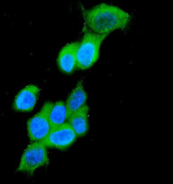
ARG40282 anti-Cytokeratin 19 antibody ICC/IF image
Immunofluorescence: MCF-7 cells were blocked with 10% goat serum and then stained with ARG40282 anti-Cytokeratin 19 antibody (green) at 2 µg/ml dilution, overnight at 4°C. DAPI (blue) for nuclear staining.
-

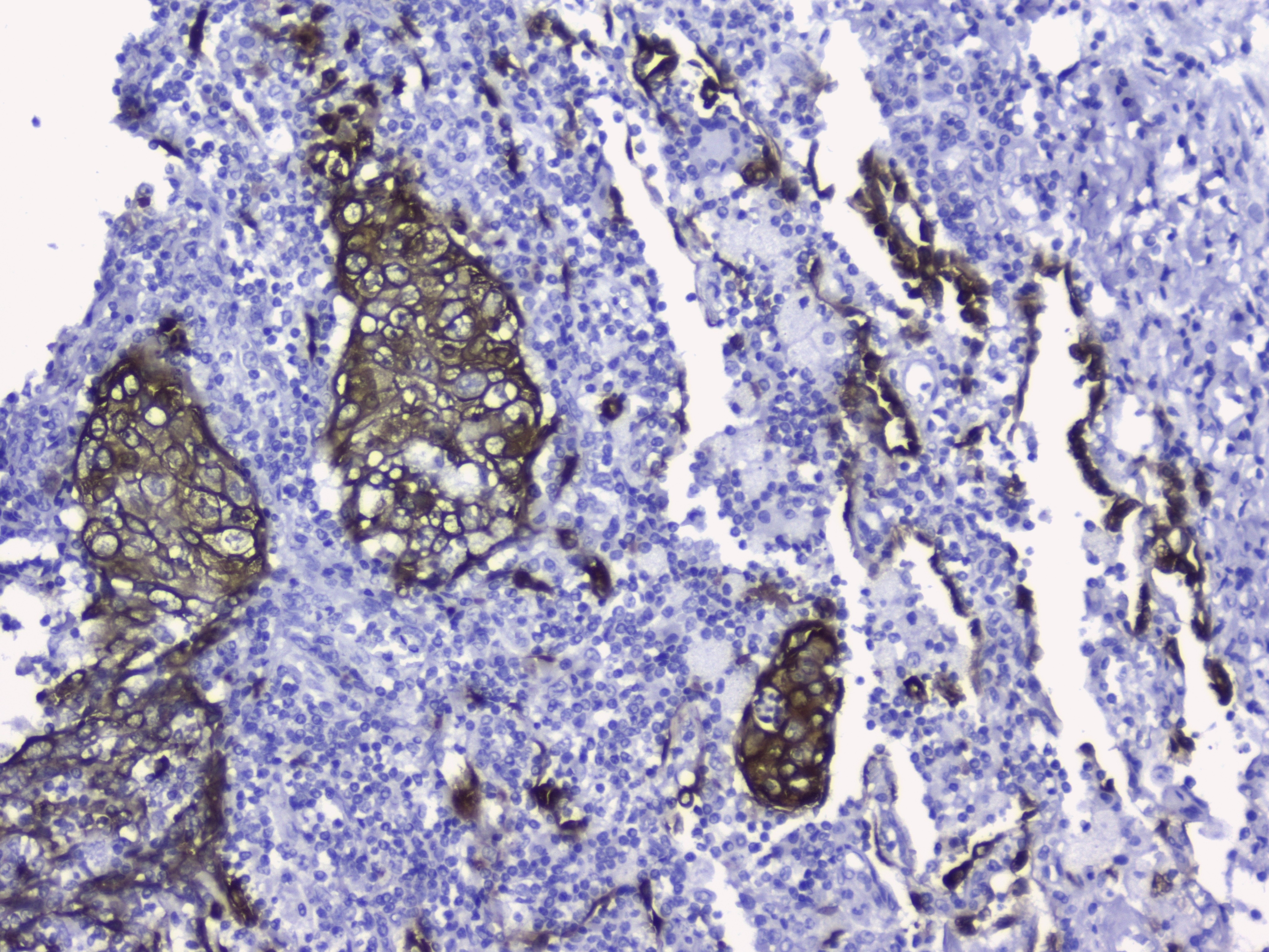
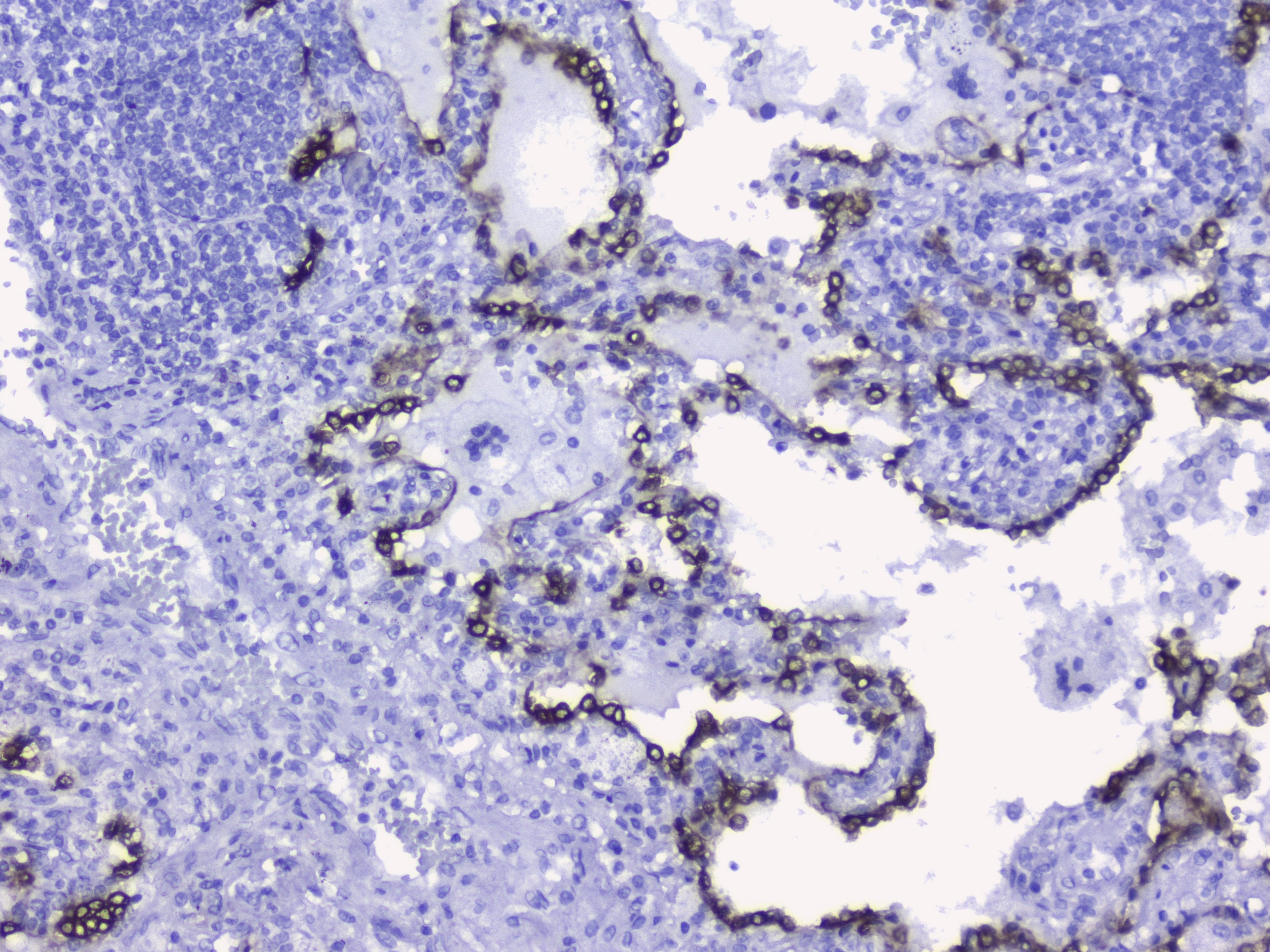
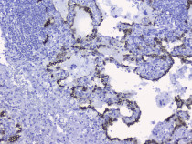
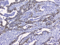
ARG40282 anti-Cytokeratin 19 antibody IHC-P image
Immunohistochemistry: Paraffin-embedded Human lung cancer tissue. Antigen Retrieval: Heat mediation was performed in Citrate buffer (pH 6.0, epitope retrieval solution) for 20 min. The tissue section was blocked with 10% goat serum. The tissue section was then stained with ARG40282 anti-Cytokeratin 19 antibody at 2 µg/ml, overnight at 4°C.
-
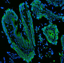
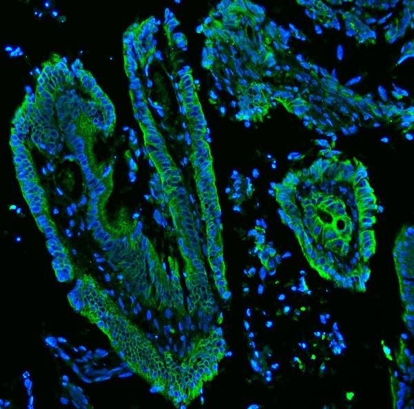
ARG40282 anti-Cytokeratin 19 antibody IHC-P image
Immunohistochemistry: Paraffin-embedded Human colon cancer tissue. Antigen Retrieval: Heat mediation was performed in EDTA buffer (pH 8.0). The tissue section was blocked with 10% goat serum. The tissue section was then stained with ARG40282 anti-Cytokeratin 19 antibody (green) at 5 µg/ml dilution, overnight at 4°C. The section was counterstained with DAPI (blue).
-
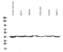
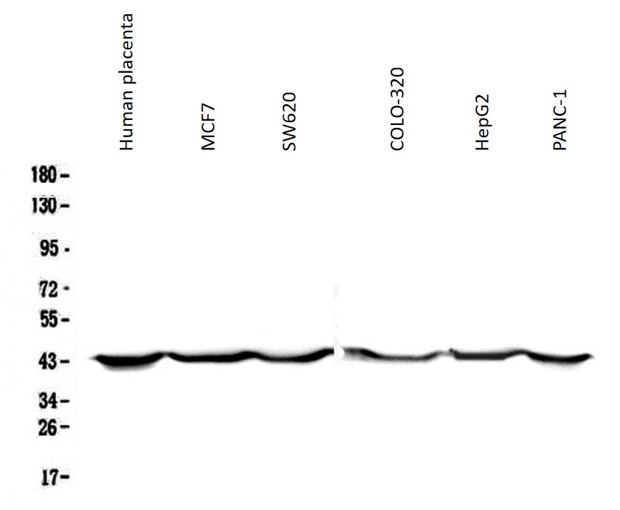
ARG40282 anti-Cytokeratin 19 antibody WB image
Western blot: 50 µg of samples under reducing conditions. Human placenta, MCF7, SW620, COLO-320, HepG2 and PANC-1 whole cell lysates stained with ARG40282 anti-Cytokeratin 19 antibody at 0.5 µg/ml, overnight at 4°C.
-
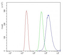
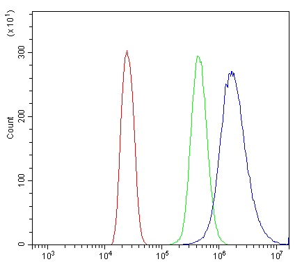
ARG40282 anti-Cytokeratin 19 antibody FACS image
Flow Cytometry: MCF-7 cells were blocked with 10% normal goat serum and then stained with ARG40282 anti-Cytokeratin 19 antibody (blue) at 1 µg/10^6 cells for 30 min at 20°C, followed by incubation with DyLight®488 labelled secondary antibody. Isotype control antibody (green) was rabbit IgG (1 µg/10^6 cells) used under the same conditions. Unlabelled sample (red) was also used as a control.
-
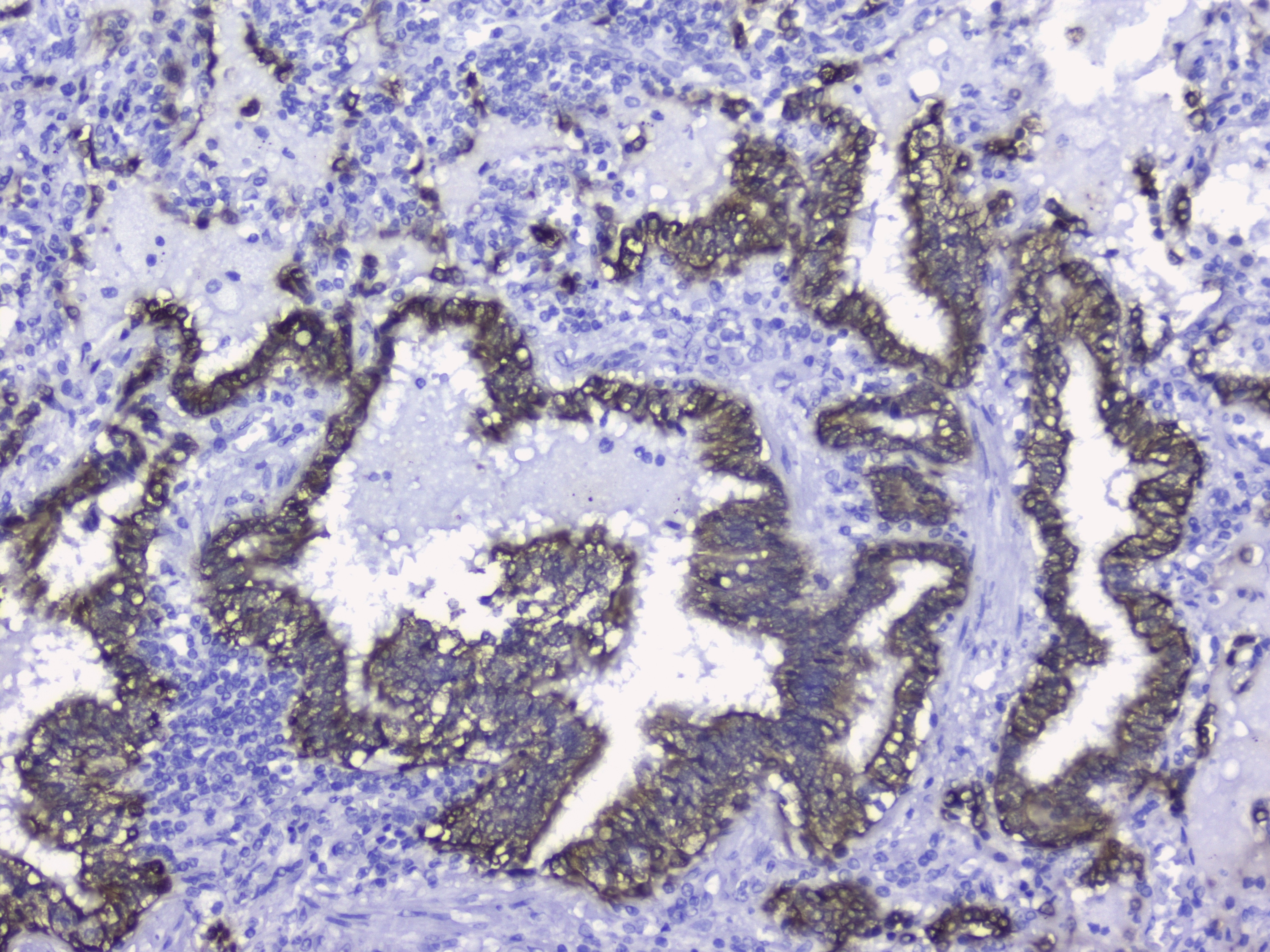
ARG40282 anti-Cytokeratin 19 antibody IHC-P image
Immunohistochemistry: Paraffin-embedded Human lung cancer tissue. Antigen Retrieval: Heat mediation was performed in Citrate buffer (pH 6.0, epitope retrieval solution) for 20 min. The tissue section was blocked with 10% goat serum. The tissue section was then stained with ARG40282 anti-Cytokeratin 19 antibody at 2 µg/ml, overnight at 4°C.
-
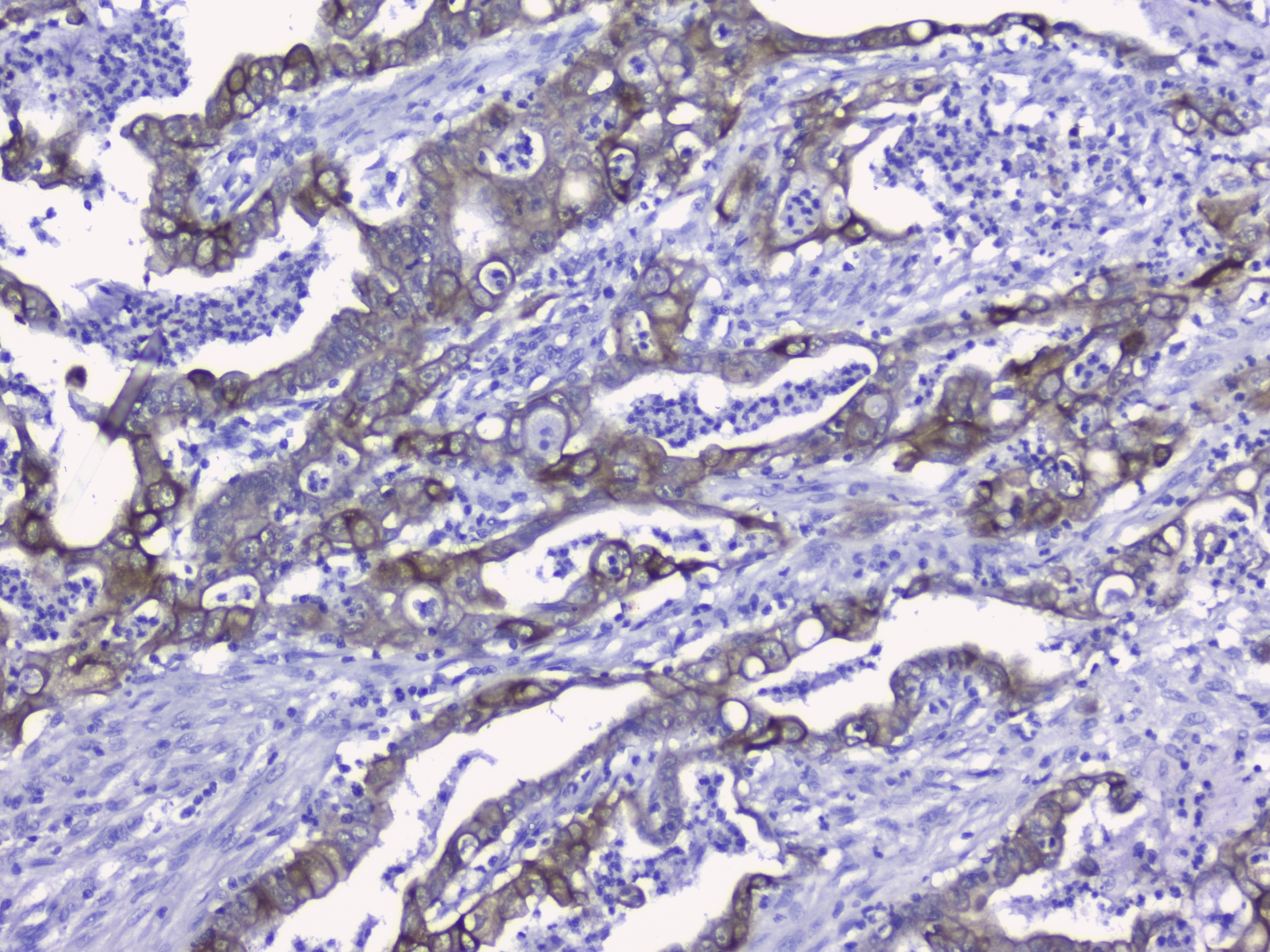
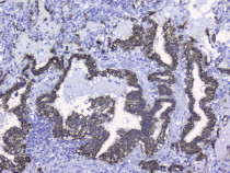
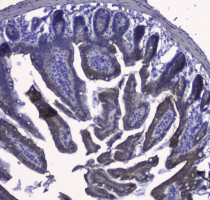
ARG40282 anti-Cytokeratin 19 antibody IHC-P image
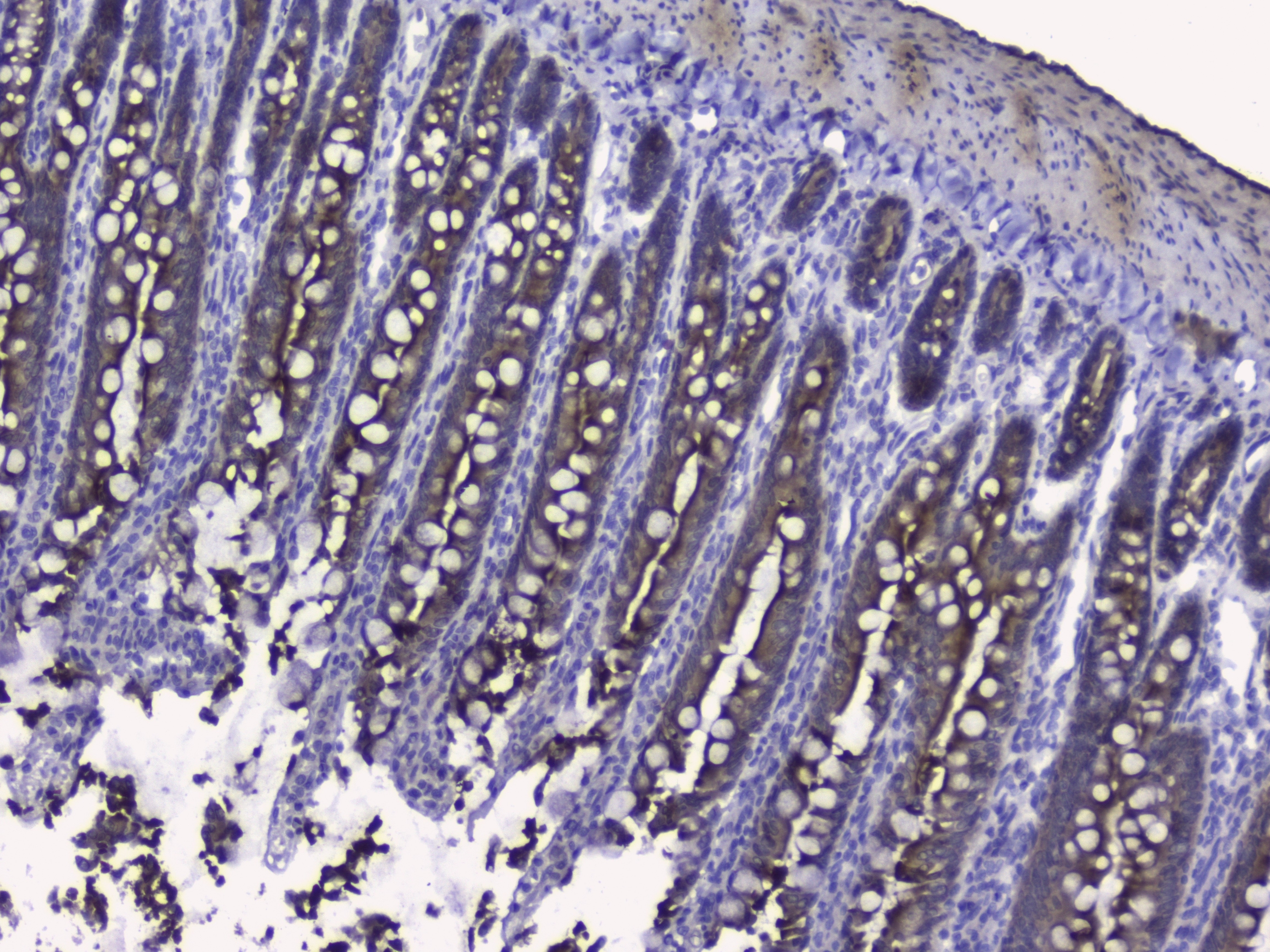
Immunohistochemistry: Paraffin-embedded Human intestinal cancer tissue. Antigen Retrieval: Heat mediation was performed in Citrate buffer (pH 6.0, epitope retrieval solution) for 20 min. The tissue section was blocked with 10% goat serum. The tissue section was then stained with ARG40282 anti-Cytokeratin 19 antibody at 2 µg/ml, overnight at 4°C.
-
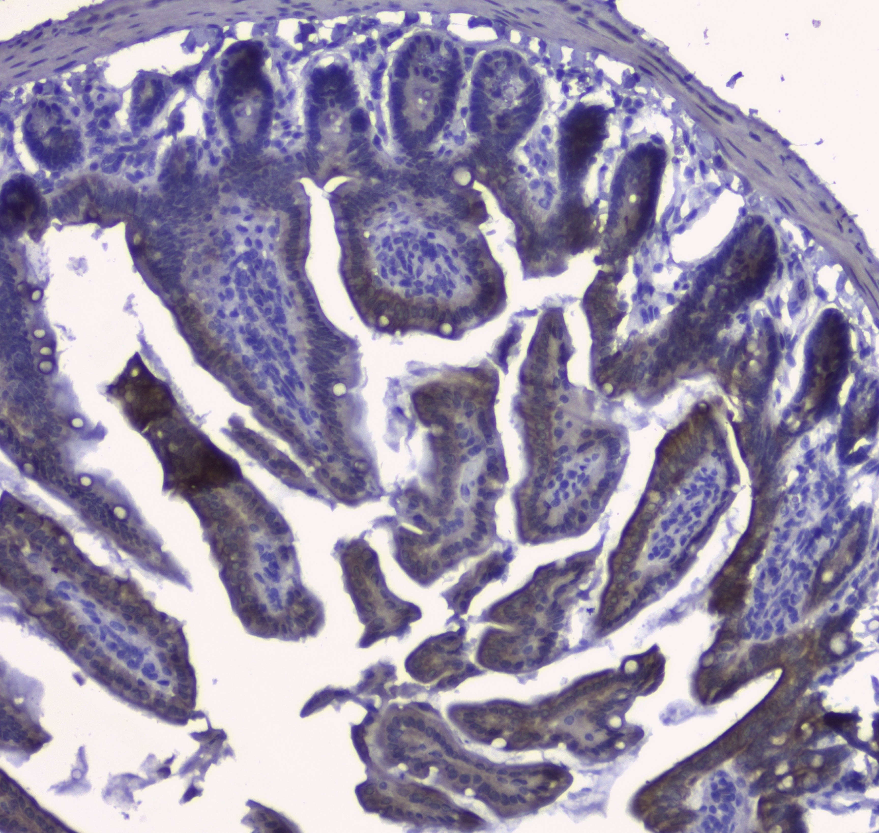
ARG40282 anti-Cytokeratin 19 antibody IHC-P image
Immunohistochemistry: Paraffin-embedded Mouse small intestine tissue. Antigen Retrieval: Heat mediation was performed in Citrate buffer (pH 6.0, epitope retrieval solution) for 20 min. The tissue section was blocked with 10% goat serum. The tissue section was then stained with ARG40282 anti-Cytokeratin 19 antibody at 2 µg/ml, overnight at 4°C.
-
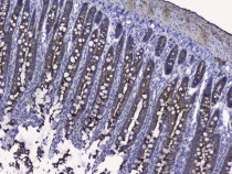
ARG40282 anti-Cytokeratin 19 antibody IHC-P image
Immunohistochemistry: Paraffin-embedded Rat small intestine tissue. Antigen Retrieval: Heat mediation was performed in Citrate buffer (pH 6.0, epitope retrieval solution) for 20 min. The tissue section was blocked with 10% goat serum. The tissue section was then stained with ARG40282 anti-Cytokeratin 19 antibody at 2 µg/ml, overnight at 4°C.
-
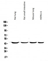
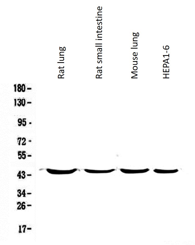
ARG40282 anti-Cytokeratin 19 antibody WB image
Western blot: 50 µg of samples under reducing conditions. Rat lung, Rat small intestine, Mouse lung and Mouse HEPA1-6 whole cell lysates stained with ARG40282 anti-Cytokeratin 19 antibody at 0.5 µg/ml, overnight at 4°C.