ARG40997
anti-DARS2 antibody
anti-DARS2 antibody for Flow cytometry,ICC/IF,IHC-Formalin-fixed paraffin-embedded sections,Western blot and Human
Overview
| Product Description | Rabbit Polyclonal antibody recognizes DARS2 |
|---|---|
| Tested Reactivity | Hu |
| Tested Application | FACS, ICC/IF, IHC-P, WB |
| Host | Rabbit |
| Clonality | Polyclonal |
| Isotype | IgG |
| Target Name | DARS2 |
| Antigen Species | Human |
| Immunogen | Recombinant protein corresponding to D334-A448 of Human DARS2. |
| Conjugation | Un-conjugated |
| Alternate Names | ASPRS; Aspartate--tRNA ligase, mitochondrial; MT-ASPRS; LBSL; AspRS; Aspartyl-tRNA synthetase; EC 6.1.1.12 |
Application Instructions
| Application Suggestion |
|
||||||||||
|---|---|---|---|---|---|---|---|---|---|---|---|
| Application Note | IHC-P: Antigen Retrieval: Heat mediation was performed in Citrate buffer (pH 6.0) for 20 min. * The dilutions indicate recommended starting dilutions and the optimal dilutions or concentrations should be determined by the scientist. |
||||||||||
| Observed Size | 74 kDa |
Properties
| Form | Liquid |
|---|---|
| Purification | Affinity purification with immunogen. |
| Buffer | 0.2% Na2HPO4, 0.9% NaCl, 0.05% Sodium azide and 4% Trehalose. |
| Preservative | 0.05% Sodium azide |
| Stabilizer | 4% Trehalose |
| Concentration | 0.5 mg/ml |
| Storage Instruction | For continuous use, store undiluted antibody at 2-8°C for up to a week. For long-term storage, aliquot and store at -20°C or below. Storage in frost free freezers is not recommended. Avoid repeated freeze/thaw cycles. Suggest spin the vial prior to opening. The antibody solution should be gently mixed before use. |
| Note | For laboratory research only, not for drug, diagnostic or other use. |
Bioinformation
| Database Links |
Swiss-port # Q6PI48 Human Aspartate--tRNA ligase, mitochondrial |
|---|---|
| Gene Symbol | DARS2 |
| Gene Full Name | aspartyl-tRNA synthetase 2, mitochondrial |
| Background | The protein encoded by this gene belongs to the class-II aminoacyl-tRNA synthetase family. It is a mitochondrial enzyme that specifically aminoacylates aspartyl-tRNA. Mutations in this gene are associated with leukoencephalopathy with brainstem and spinal cord involvement and lactate elevation (LBSL). [provided by RefSeq, Nov 2009] |
| Cellular Localization | Mitochondrion matrix. [UniProt] |
| Calculated MW | 74 kDa |
Images (7) Click the Picture to Zoom In
-

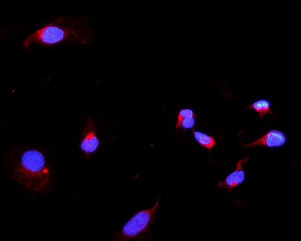
ARG40997 anti-DARS2 antibody ICC/IF image
Immunofluorescence: U2OS cells were blocked with 10% goat serum and then stained with ARG40997 anti-DARS2 antibody (red) at 2 µg/ml dilution, overnight at 4°C. DAPI (blue) for nuclear staining.
-
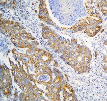
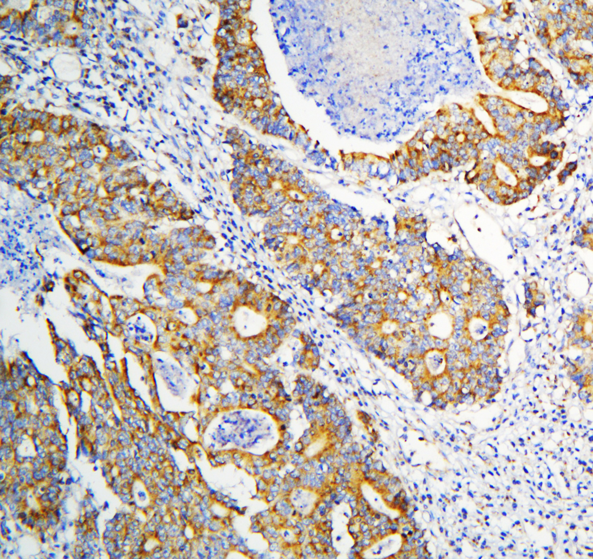
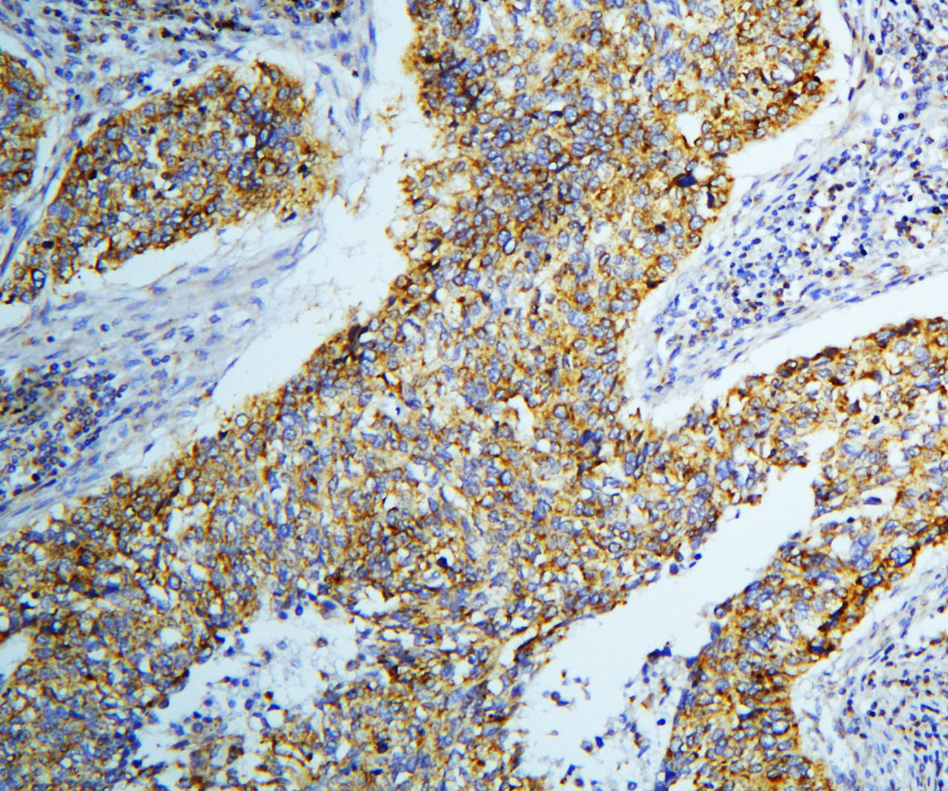
ARG40997 anti-DARS2 antibody IHC-P image
Immunohistochemistry: Paraffin-embedded Human intestinal cancer tissue. Antigen Retrieval: Heat mediation was performed in Citrate buffer (pH 6.0, epitope retrieval solution) for 20 min. The tissue section was blocked with 10% goat serum. The tissue section was then stained with ARG40997 anti-DARS2 antibody at 1 µg/ml dilution, overnight at 4°C.
-
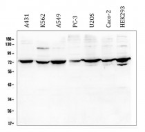
ARG40997 anti-DARS2 antibody WB image
Western blot: 50 µg of samples under reducing conditions. A431, K562, A549, PC-3, U2OS, Caco-2 and HEK293 whole cell lysates stained with ARG40997 anti-DARS2 antibody at 0.5 µg/ml, overnight at 4°C.
-
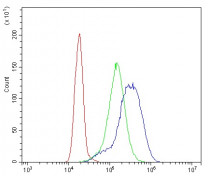
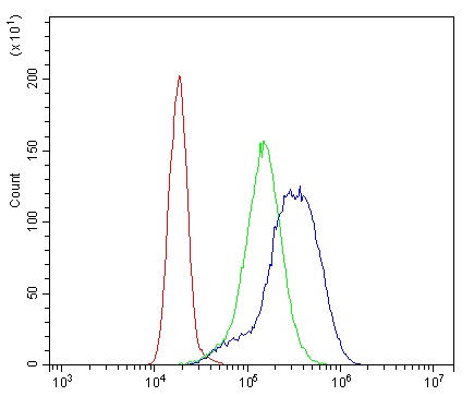
ARG40997 anti-DARS2 antibody FACS image
Flow Cytometry: K562 cells were blocked with 10% normal goat serum and then stained with ARG40997 anti-DARS2 antibody (blue) at 1 µg/10^6 cells for 30 min at 20°C, followed by incubation with DyLight®488 labelled secondary antibody. Isotype control antibody (green) was rabbit IgG (1 µg/10^6 cells) used under the same conditions. Unlabelled sample (red) was also used as a control.
-
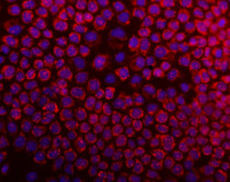
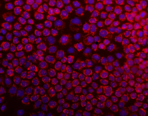
ARG40997 anti-DARS2 antibody ICC/IF image
Immunofluorescence: A431 cells were blocked with 10% goat serum and then stained with ARG40997 anti-DARS2 antibody (red) at 2 µg/ml dilution, overnight at 4°C. DAPI (blue) for nuclear staining.
-
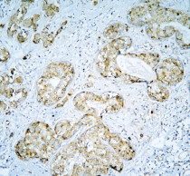
ARG40997 anti-DARS2 antibody IHC-P image
Immunohistochemistry: Paraffin-embedded Human mammary cancer tissue. Antigen Retrieval: Heat mediation was performed in Citrate buffer (pH 6.0, epitope retrieval solution) for 20 min. The tissue section was blocked with 10% goat serum. The tissue section was then stained with ARG40997 anti-DARS2 antibody at 1 µg/ml dilution, overnight at 4°C.
-
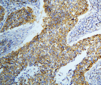
ARG40997 anti-DARS2 antibody IHC-P image
Immunohistochemistry: Paraffin-embedded Human lung cancer tissue. Antigen Retrieval: Heat mediation was performed in Citrate buffer (pH 6.0, epitope retrieval solution) for 20 min. The tissue section was blocked with 10% goat serum. The tissue section was then stained with ARG40997 anti-DARS2 antibody at 1 µg/ml dilution, overnight at 4°C.