ARG59736
anti-DENR antibody [1542CT106.51.79]
anti-DENR antibody [1542CT106.51.79] for Flow cytometry,ICC/IF,IHC-Formalin-fixed paraffin-embedded sections,Western blot and Human
Overview
| Product Description | Mouse Monoclonal antibody recognizes DENR |
|---|---|
| Tested Reactivity | Hu |
| Tested Application | FACS, ICC/IF, IHC-P, WB |
| Host | Mouse |
| Clonality | Monoclonal |
| Clone | 1542CT106.51.79 |
| Isotype | IgG2b, kappa |
| Target Name | DENR |
| Antigen Species | Human |
| Immunogen | Recombinant protein of Human DENR. |
| Conjugation | Un-conjugated |
| Alternate Names | DRP; DRP1; Smooth muscle cell-associated protein 3; Protein DRP1; Density-regulated protein; SMAP-3 |
Application Instructions
| Application Suggestion |
|
||||||||||
|---|---|---|---|---|---|---|---|---|---|---|---|
| Application Note | IHC-P: Antigen Retrieval: Heat mediation was performed in Citrate buffer (pH 6.0). * The dilutions indicate recommended starting dilutions and the optimal dilutions or concentrations should be determined by the scientist. |
||||||||||
| Positive Control | A431 |
Properties
| Form | Liquid |
|---|---|
| Purification | Purification with Protein G. |
| Buffer | PBS and 0.09% (W/V) Sodium azide. |
| Preservative | 0.09% (W/V) Sodium azide. |
| Storage Instruction | For continuous use, store undiluted antibody at 2-8°C for up to a week. For long-term storage, aliquot and store at -20°C or below. Storage in frost free freezers is not recommended. Avoid repeated freeze/thaw cycles. Suggest spin the vial prior to opening. The antibody solution should be gently mixed before use. |
| Note | For laboratory research only, not for drug, diagnostic or other use. |
Bioinformation
| Database Links | |
|---|---|
| Gene Symbol | DENR |
| Gene Full Name | density-regulated protein |
| Background | This gene encodes a protein whose expression was found to increase in cultured cells at high density but not during growth arrest. This gene was also shown to have increased expression in cells overexpressing HER-2/neu proto-oncogene. The protein contains an SUI1 domain. In budding yeast, SUI1 is a translation initiation factor that along with eIF-2 and the initiator tRNA-Met, directs the ribosome to the proper translation start site. Proteins similar to SUI have been found in mammals, insects, and plants. [provided by RefSeq, Jul 2008] |
| Function | May be involved in the translation of target mRNAs by scanning and recognition of the initiation codon. Involved in translation initiation; promotes recruitmnet of aminoacetyled initiator tRNA to P site of 40S ribosomes. Can promote release of deacylated tRNA and mRNA from recycled 40S subunits following ABCE1-mediated dissociation of post-termination ribosomal complexes into subunits. Plays a role in the modulation of the translational profile of a subset of cancer-related mRNAs when recruited to the translational initiation complex by the oncogene MCTS1. [UniProt] |
| Calculated MW | 22 kDa |
Images (4) Click the Picture to Zoom In
-

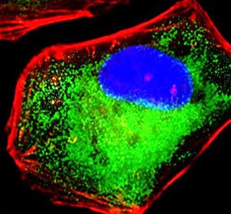
ARG59736 anti-DENR antibody ICC/IF image
Immunofluorescence: 4% paraformaldehyde-fixed, 0.1% Triton X-100 permeabilized HeLa cells stained with ARG59736 anti-DENR antibody (green) at 1:25 dilution. Cytoplasmic actin is detected with Dylight® 554 Phalloidin (red) at 1:100 dilution. The nuclear counter stain is DAPI (blue).
-
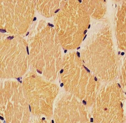
ARG59736 anti-DENR antibody IHC-P image
Immunohistochemistry: Paraformaldehyde-fixed and paraffin-embedded Human skeletal muscle. Tissue was blocked with 3% BSA for 0.5 hour at RT. Antigen Retrieval: Heat mediation was performed in Citrate buffer (pH 6.0). Samples were stained with ARG59736 anti-DENR antibody at 1:25 for 1 hours at 37°C.
-

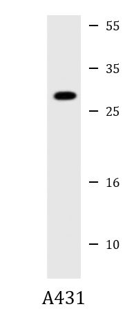
ARG59736 anti-DENR antibody WB image
Western blot: 20 µg of A431 cell lysate stained with ARG59736 anti-DENR antibody at 1:1000 - 1:2000 dilution.
-
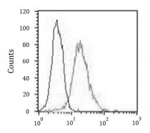
ARG59736 anti-DENR antibody FACS image
Flow Cytometry: HeLa cells were fixed with 2% paraformaldehyde (10 min) and then permeabilized with 90% methanol for 10 min. Cells were then incubated in 2% BSA to block non-specific protein-protein interactions followed by ARG59736 anti-DENR antibody (right histogram) at 1:25 dilution for 60 min at 37°C, followed by DyLight®488 labelled secondary antibody. Isotype control antibody (left histogram) was Mouse IgG2b (1 µg/10^6 cells) used under the same conditions. Acquisition of > 10000 events was performed.