ARG65204
anti-DYDC1 antibody
anti-DYDC1 antibody for IHC-Formalin-fixed paraffin-embedded sections,Western blot and Human,Mouse,Rat
Cell Biology and Cellular Response antibody
Overview
| Product Description | Goat Polyclonal antibody recognizes DYDC1 |
|---|---|
| Tested Reactivity | Hu, Ms, Rat |
| Predict Reactivity | Cow |
| Tested Application | IHC-P, WB |
| Host | Goat |
| Clonality | Polyclonal |
| Isotype | IgG |
| Target Name | DYDC1 |
| Antigen Species | Human |
| Immunogen | C-TLAEISDRYGAPN |
| Conjugation | Un-conjugated |
| Alternate Names | DPY30 domain-containing protein 1; DPY30D1 |
Application Instructions
| Application Suggestion |
|
||||||
|---|---|---|---|---|---|---|---|
| Application Note | WB: Recommend incubate at RT for 1h. IHC-P: Antigen Retrieval: Steam tissue section in Citrate buffer (pH 6.0). * The dilutions indicate recommended starting dilutions and the optimal dilutions or concentrations should be determined by the scientist. |
Properties
| Form | Liquid |
|---|---|
| Purification | Purified from goat serum by antigen affinity chromatography. |
| Buffer | Tris saline (pH 7.3), 0.02% Sodium azide and 0.5% BSA. |
| Preservative | 0.02% Sodium azide |
| Stabilizer | 0.5% BSA |
| Concentration | 0.5 mg/ml |
| Storage Instruction | For continuous use, store undiluted antibody at 2-8°C for up to a week. For long-term storage, aliquot and store at -20°C or below. Storage in frost free freezers is not recommended. Avoid repeated freeze/thaw cycles. Suggest spin the vial prior to opening. The antibody solution should be gently mixed before use. |
| Note | For laboratory research only, not for drug, diagnostic or other use. |
Bioinformation
| Database Links | |
|---|---|
| Background | This gene encodes a member of a family of proteins that contains a DPY30 domain. The encoded protein is involved in acrosome formation during spermatid development. This gene locus overlaps with a closely related gene on the opposite strand. Alternative splicing results in multiple transcript variants. [provided by RefSeq, Jun 2012] |
| Research Area | Cell Biology and Cellular Response antibody |
| Calculated MW | 21 kDa |
Images (2) Click the Picture to Zoom In
-

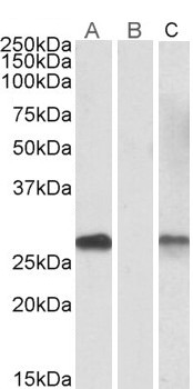
ARG65204 anti-DYDC1 antibody WB image
Western Blot: Lane A - HEK293 overexpressing Human DYDC1 lysate (10µg protein in RIPA buffer) stained with ARG65204 anti-DYDC1 (aa142-154) antibody (1µg/ml); Lane B - HEK293 mock-transfected lysate (10µg protein in RIPA buffer) stained with ARG65204 anti-DYDC1 (aa142-154) antibody (0.1µg/ml . Lane C - anti-MYC Tag (1/1000) staining HEK293 overexpressing Human DYDC1 lysate (10µg protein in RIPA buffer). Primary incubations were for 1 hour. Detected by chemiluminescence.
-

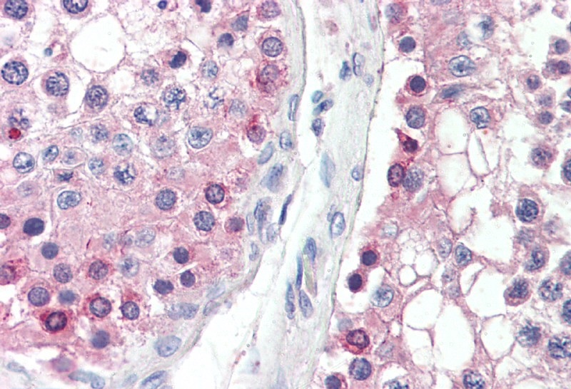
ARG65204 anti-DYDC1 antibody IHC-P image
Immunohistochemistry: Paraffin-embedded Human testis tissue. Antigen Retrieval: Steam tissue section in Citrate buffer (pH 6.0). The tissue section was stained with ARG65204 anti-DYDC1 antibody at 5 µg/ml dilution followed by AP-staining.