ARG63341
anti-FOXC1 antibody
anti-FOXC1 antibody for Flow cytometry,ICC/IF,IHC-Formalin-fixed paraffin-embedded sections and Human
Developmental Biology antibody; Gene Regulation antibody; Chondrogenesis Study antibody
Overview
| Product Description | Goat Polyclonal antibody recognizes FOXC1 |
|---|---|
| Tested Reactivity | Hu |
| Predict Reactivity | Ms, Rat |
| Tested Application | FACS, ICC/IF, IHC-P |
| Host | Goat |
| Clonality | Polyclonal |
| Isotype | IgG |
| Target Name | FOXC1 |
| Antigen Species | Human |
| Immunogen | RTSGAFVYDCSKF |
| Conjugation | Un-conjugated |
| Alternate Names | IRID1; Forkhead box protein C1; FREAC3; IHG1; ARA; IGDA; FREAC-3; Forkhead-related transcription factor 3; RIEG3; FKHL7; Forkhead-related protein FKHL7 |
Application Instructions
| Application Suggestion |
|
||||||||
|---|---|---|---|---|---|---|---|---|---|
| Application Note | IHC-P: Antigen Retrieval: Steam tissue section in Citrate buffer (pH 6.0). * The dilutions indicate recommended starting dilutions and the optimal dilutions or concentrations should be determined by the scientist. |
Properties
| Form | Liquid |
|---|---|
| Purification | Purified from goat serum by antigen affinity chromatography. |
| Buffer | Tris saline (pH 7.3), 0.02% Sodium azide and 0.5% BSA. |
| Preservative | 0.02% Sodium azide |
| Stabilizer | 0.5% BSA |
| Concentration | 0.5 mg/ml |
| Storage Instruction | For continuous use, store undiluted antibody at 2-8°C for up to a week. For long-term storage, aliquot and store at -20°C or below. Storage in frost free freezers is not recommended. Avoid repeated freeze/thaw cycles. Suggest spin the vial prior to opening. The antibody solution should be gently mixed before use. |
| Note | For laboratory research only, not for drug, diagnostic or other use. |
Bioinformation
| Database Links | |
|---|---|
| Background | This gene belongs to the forkhead family of transcription factors which is characterized by a distinct DNA-binding forkhead domain. The specific function of this gene has not yet been determined; however, it has been shown to play a role in the regulation of embryonic and ocular development. Mutations in this gene cause various glaucoma phenotypes including primary congenital glaucoma, autosomal dominant iridogoniodysgenesis anomaly, and Axenfeld-Rieger anomaly. [provided by RefSeq, Jul 2008] |
| Highlight | Related Antibody Duos and Panels: ARG30314 Chondrogenesis Marker Antibody Panel Related products: FOXC1 antibodies; FOXC1 Duos / Panels; Anti-Goat IgG secondary antibodies; |
| Research Area | Developmental Biology antibody; Gene Regulation antibody; Chondrogenesis Study antibody |
| Calculated MW | 57 kDa |
| PTM | Phosphorylated. |
Images (5) Click the Picture to Zoom In
-

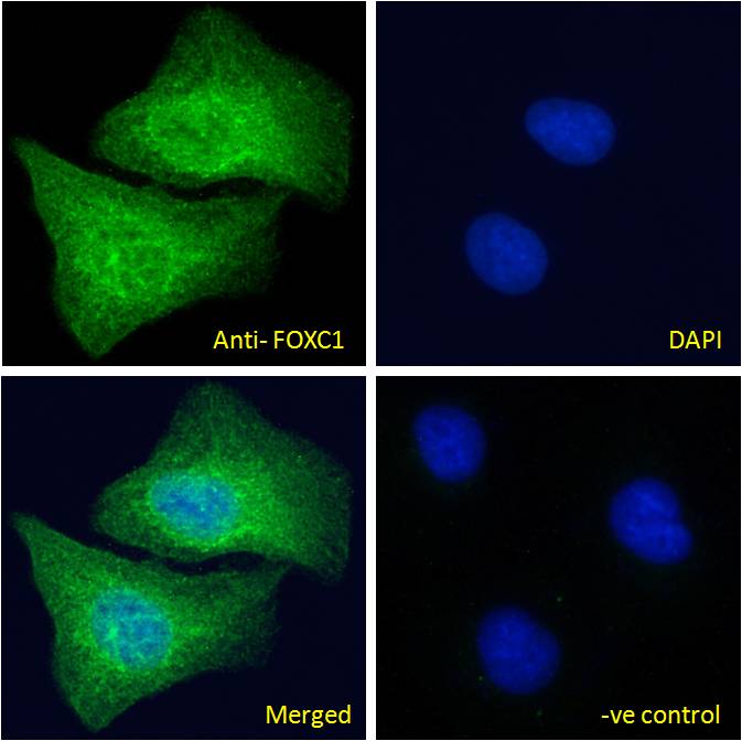
ARG63341 anti-FOXC1 antibody ICC/IF image
Immunofluorescence: Paraformaldehyde fixed U2OS cells permeabilized with 0.15% Triton. Cells were stained with ARG63341 anti-FOXC1 antibody (green) at 10 µg/ml dilution for 1 hour. DAPI (blue) for nuclear staining. Negative control: Unimmunized goat IgG (green) at 10 µg/ml dilution.
-
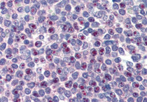
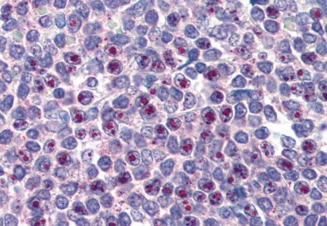
ARG63341 anti-FOXC1 antibody IHC-P image
Immunohistochemistry: Paraffin-embedded Human spleen tissue. Antigen Retrieval: Steam tissue section in Citrate buffer (pH 6.0). The tissue section was stained with ARG63341 anti-FOXC1 antibody at 3.75 µg/ml dilution followed by AP-staining.
-
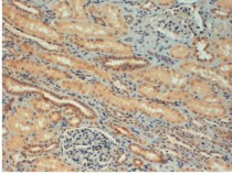
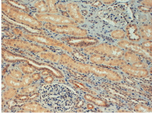
ARG63341 anti-FOXC1 antibody IHC-P image
Immunohistochemistry: paraffin embedded Human Kidney. (Steamed antigen retrieval with citrate buffer pH 6) stained with ARG63341 anti-FOXC1 antibody at 4 µg/ml dilution followed by HRP-staining.
-
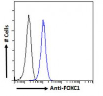
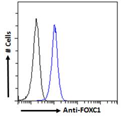
ARG63341 anti-FOXC1 antibody FACS image
Flow Cytometry: Paraformaldehyde-fixed HEK293 cells permeabilized with 0.5% Triton. Cells were stained with ARG63341 anti-FOXC1 antibody (blue line) at 10 µg/ml dilution for 1 hour, followed by incubation with Alexa FluorR 488 labelled secondary antibody. IgG control: Unimmunized goat IgG (black line).
-
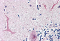
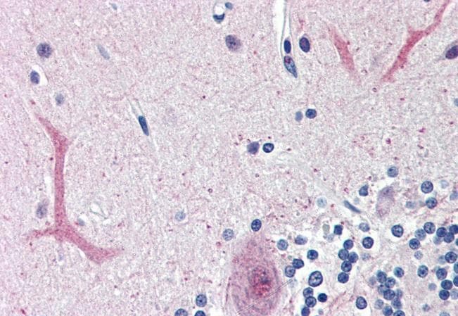
ARG63341 anti-FOXC1 antibody IHC-P image
Immunohistochemistry: Paraffin-embedded Human cerebellum tissue. Antigen Retrieval: Steam tissue section in Citrate buffer (pH 6.0). The tissue section was stained with ARG63341 anti-FOXC1 antibody at 3.75 µg/ml dilution followed by AP-staining.