ARG58791
anti-HGD antibody
anti-HGD antibody for Flow cytometry,ICC/IF,IHC-Formalin-fixed paraffin-embedded sections,Western blot and Human,Mouse,Rat
Overview
| Product Description | Rabbit Polyclonal antibody recognizes HGD |
|---|---|
| Tested Reactivity | Hu, Ms, Rat |
| Tested Application | FACS, ICC/IF, IHC-P, WB |
| Host | Rabbit |
| Clonality | Polyclonal |
| Isotype | IgG |
| Target Name | HGD |
| Antigen Species | Human |
| Immunogen | Recombinant protein corresponding to D374-N445 of Human HGD. |
| Conjugation | Un-conjugated |
| Alternate Names | EC 1.13.11.5; AKU; HGO; Homogentisate 1,2-dioxygenase; Homogentisate oxygenase; Homogentisicase; Homogentisic acid oxidase |
Application Instructions
| Application Suggestion |
|
||||||||||
|---|---|---|---|---|---|---|---|---|---|---|---|
| Application Note | IHC-P: Antigen Retrieval: Heat mediation was performed in Citrate buffer (pH 6.0) for 20 min. * The dilutions indicate recommended starting dilutions and the optimal dilutions or concentrations should be determined by the scientist. |
Properties
| Form | Liquid |
|---|---|
| Purification | Affinity purification with immunogen. |
| Buffer | 0.9% NaCl, 0.2% Na2HPO4, 0.05% Sodium azide and 4% Trehalose. |
| Preservative | 0.05% Sodium azide |
| Stabilizer | 4% Trehalose |
| Concentration | 0.5 mg/ml |
| Storage Instruction | For continuous use, store undiluted antibody at 2-8°C for up to a week. For long-term storage, aliquot and store at -20°C or below. Storage in frost free freezers is not recommended. Avoid repeated freeze/thaw cycles. Suggest spin the vial prior to opening. The antibody solution should be gently mixed before use. |
| Note | For laboratory research only, not for drug, diagnostic or other use. |
Bioinformation
| Database Links | |
|---|---|
| Gene Symbol | HGD |
| Gene Full Name | homogentisate 1,2-dioxygenase |
| Background | This gene encodes the enzyme homogentisate 1,2 dioxygenase. This enzyme is involved in the catabolism of the amino acids tyrosine and phenylalanine. Mutations in this gene are the cause of the autosomal recessive metabolism disorder alkaptonuria.[provided by RefSeq, May 2010] |
| Calculated MW | 50 kDa |
Images (5) Click the Picture to Zoom In
-
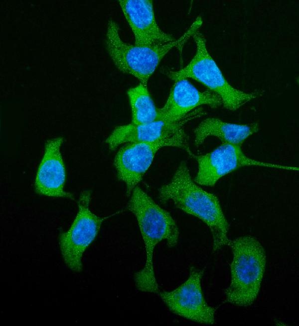
ARG58791 anti-HGD antibody ICC/IF image
Immunofluorescence: Caco-2 cells were blocked with 10% goat serum and then stained with ARG58791 anti-HGD antibody (green) at 5 µg/ml dilution, overnight at 4°C. DAPI (blue) for nuclear staining.
-
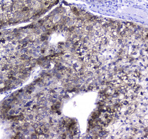
ARG58791 anti-HGD antibody IHC-P image
Immunohistochemistry: Paraffin-embedded Human liver cancer tissue. Antigen Retrieval: Heat mediated was performed in Citrate buffer (pH 6.0, epitope retrieval solution) for 20 min. The tissue section was blocked with 10% goat serum. The tissue section was then stained with ARG58791 anti-HGD antibody at 2 µg/ml, overnight at 4°C.
-
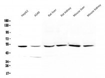
ARG58791 anti-HGD antibody WB image
Western blot: 50 µg of samples under reducing conditions. HepG2, A549, Rat liver, Rat kidney, Mouse liver and Mouse kidney lysates stained with ARG58791 anti-HGD antibody at 0.5 µg/ml, overnight at 4°C.
-
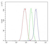
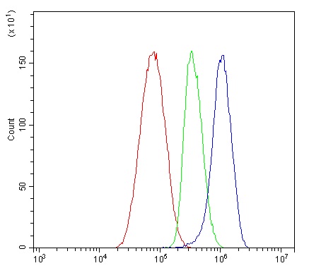
ARG58791 anti-HGD antibody FACS image
Flow Cytometry: HepG2 cells were blocked with 10% normal goat serum and then stained with ARG58791 anti-HGD antibody (blue) at 1 µg/10^6 cells for 30 min at 20°C, followed by incubation with DyLight®488 labelled secondary antibody. Isotype control antibody (green) was rabbit IgG (1 µg/10^6 cells) used under the same conditions. Unlabelled sample (red) was also used as a control.
-

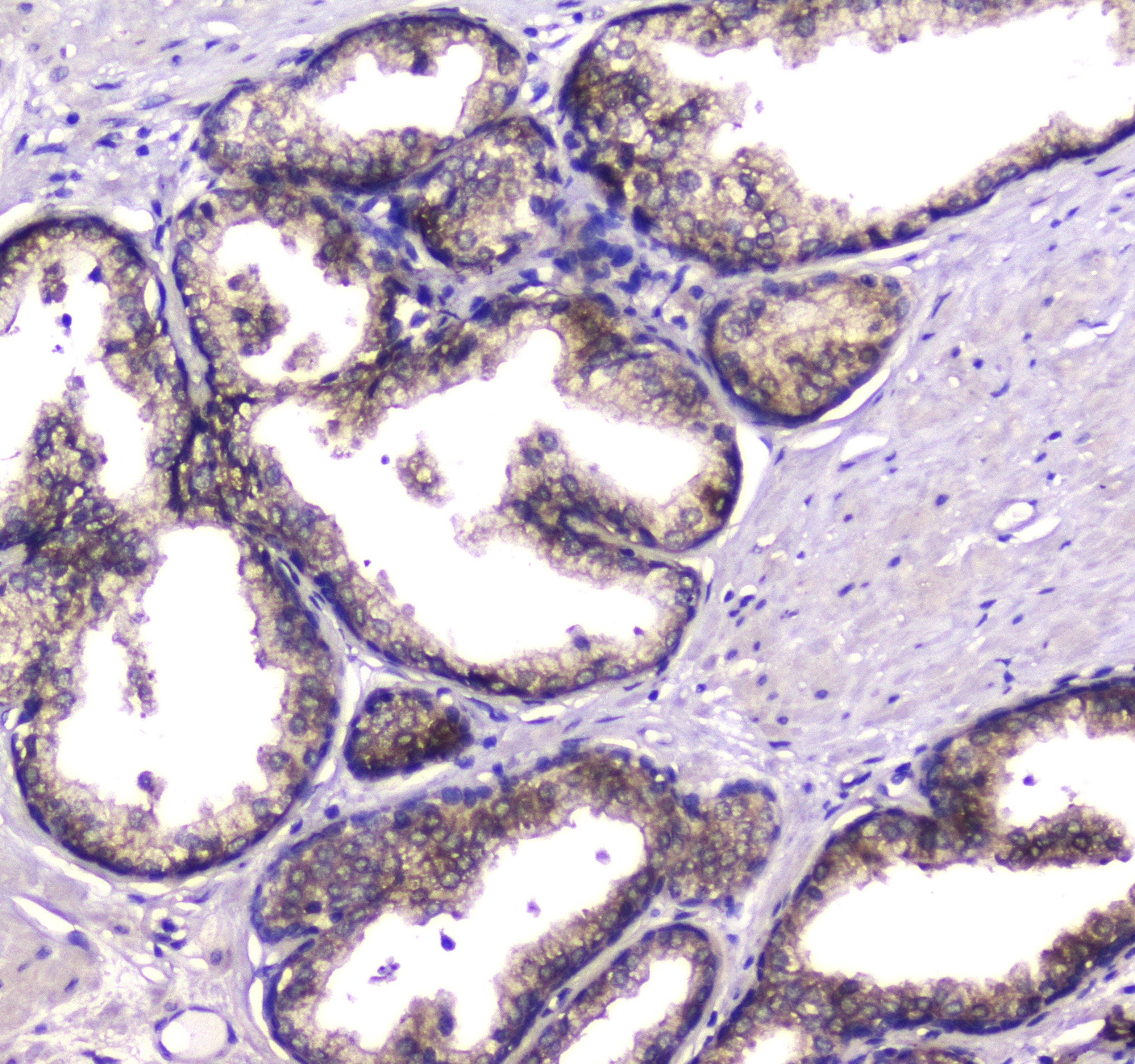
ARG58791 anti-HGD antibody IHC-P image
Immunohistochemistry: Paraffin-embedded Human prostatic cancer tissue. Antigen Retrieval: Heat mediated was performed in Citrate buffer (pH 6.0, epitope retrieval solution) for 20 min. The tissue section was blocked with 10% goat serum. The tissue section was then stained with ARG58791 anti-HGD antibody at 2 µg/ml, overnight at 4°C.