ARG66663
anti-JAK1 antibody
anti-JAK1 antibody for IHC-Formalin-fixed paraffin-embedded sections,Western blot and Human,Mouse,Rat
Overview
| Product Description | Rabbit Polyclonal antibody recognizes JAK1 |
|---|---|
| Tested Reactivity | Hu, Ms, Rat |
| Tested Application | IHC-P, WB |
| Host | Rabbit |
| Clonality | Polyclonal |
| Isotype | IgG |
| Target Name | JAK1 |
| Antigen Species | Human |
| Immunogen | Synthetic peptide corresponding to aa. 988-1037 of Human JAK1. |
| Conjugation | Un-conjugated |
| Alternate Names | JTK3; Janus kinase 1; JAK-1; Tyrosine-protein kinase JAK1; JAK1A; JAK1B; EC 2.7.10.2 |
Application Instructions
| Application Suggestion |
|
||||||
|---|---|---|---|---|---|---|---|
| Application Note | IHC-P: Antigen Retrieval: Heat mediation was performed in Sodium citrate buffer (pH 6.0) for 20 min. * The dilutions indicate recommended starting dilutions and the optimal dilutions or concentrations should be determined by the scientist. |
||||||
| Observed Size | ~ 130 kDa |
Properties
| Form | Liquid |
|---|---|
| Purification | Affinity purification with immunogen. |
| Buffer | PBS, 0.02% Sodium azide, 50% Glycerol and 0.5% BSA. |
| Preservative | 0.02% Sodium azide |
| Stabilizer | 50% Glycerol and 0.5% BSA |
| Concentration | 1 mg/ml |
| Storage Instruction | For continuous use, store undiluted antibody at 2-8°C for up to a week. For long-term storage, aliquot and store at -20°C. Storage in frost free freezers is not recommended. Avoid repeated freeze/thaw cycles. Suggest spin the vial prior to opening. The antibody solution should be gently mixed before use. |
| Note | For laboratory research only, not for drug, diagnostic or other use. |
Bioinformation
| Database Links | |
|---|---|
| Gene Symbol | JAK1 |
| Gene Full Name | Janus kinase 1 |
| Background | Janus kinase 1 (JAK1), is a member of a new class of protein-tyrosine kinases (PTK) characterized by the presence of a second phosphotransferase-related domain immediately N-terminal to the PTK domain. The second phosphotransferase domain bears all the hallmarks of a protein kinase, although its structure differs significantly from that of the PTK and threonine/serine kinase family members. JAK1 is a large, widely expressed membrane-associated phosphoprotein. JAK1 is involved in the interferon-alpha/beta and -gamma signal transduction pathways. The reciprocal interdependence between JAK1 and TYK2 activities in the interferon-alpha pathway, and between JAK1 and JAK2 in the interferon-gamma pathway, may reflect a requirement for these kinases in the correct assembly of interferon receptor complexes. These kinases couple cytokine ligand binding to tyrosine phosphorylation of various known signaling proteins and of a unique family of transcription factors termed the signal transducers and activators of transcription, or STATs. [provided by RefSeq, Jul 2008] |
| Function | Tyrosine kinase of the non-receptor type, involved in the IFN-alpha/beta/gamma signal pathway. Kinase partner for the interleukin (IL)-2 receptor. [UniProt] |
| Cellular Localization | Endomembrane system; Peripheral membrane protein. Note=Wholly intracellular, possibly membrane associated. [UniProt] |
| Calculated MW | 133 kDa |
| PTM | Autophosphorylated (PubMed:7615558). Phosphorylated on tyrosine residues in response to interferon gamma signaling (PubMed:7615558). Dephosphorylation of Tyr-1034 and Tyr-1035 by PTPN2 negatively regulates cytokine-mediated signaling (PubMed:11909529). Ubiquitinated by RNF125; leading to its degradation by the proteasome. [UniProt] |
Images (4) Click the Picture to Zoom In
-
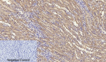
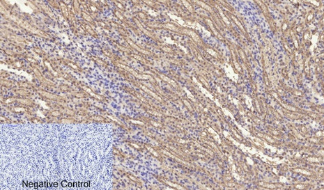
ARG66663 anti-JAK1 antibody IHC-P image
Immunohistochemistry: Paraffin-embedded Mouse kidney tissue. Antigen Retrieval: Heat mediation was performed in Sodium citrate buffer (pH 6.0) for 20 min. The tissue section was stained with ARG66663 anti-JAK1 antibody at 1:200 dilution, overnight at 4°C. Negative control was used by secondary antibody only.
-
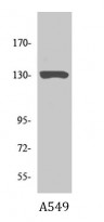
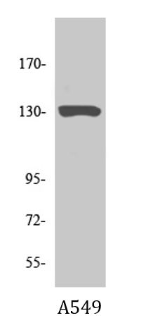
ARG66663 anti-JAK1 antibody WB image
Western blot: A549 cell lysate stained with ARG66663 anti-JAK1 antibody at 1:1000 dilution.
-
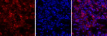
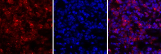
ARG66663 anti-JAK1 antibody IHC image
Immunohistochemistry: Rat lung tissue stained with ARG66663 anti-JAK1 antibody (orange-red) at 1:200 dilution, overnight at 4°C. DAPI (blue) for nuclear staining.
-

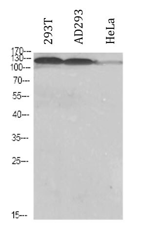
ARG66663 anti-JAK1 antibody WB image
Western blot: 293T, AD293 and HeLa cell lysates stained with ARG66663 anti-JAK1 antibody at 1:1000 dilution.