ARG66008
anti-M-CSF antibody
anti-M-CSF antibody for ELISA,IHC-Formalin-fixed paraffin-embedded sections,Neutralizing,Western blot and Human
Overview
| Product Description | Rabbit Polyclonal antibody recognizes M-CSF |
|---|---|
| Tested Reactivity | Hu |
| Tested Application | ELISA, IHC-P, Neut, WB |
| Host | Rabbit |
| Clonality | Polyclonal |
| Isotype | IgG |
| Target Name | M-CSF |
| Antigen Species | Human |
| Immunogen | E. coli derived recombinant Human M-CSF. (MEEVSEYCSH MIGSGHLQSL QRLIDSQMET SCQITFEFVD QEQLKDPVCY LKKAFLLVQD IMEDTMRFRD NTPNAIAIVQ LQELSLRLKS CFTKDYEEHD KACVRTFYET PLQLLEKVKN VFNETKNLLD KDWNIFSKNC NNSFAECSSQ GHERQSEGS) |
| Conjugation | Un-conjugated |
| Alternate Names | Macrophage colony-stimulating factor 1; CSF-1; Lanimostim; M-CSF; MCSF |
Application Instructions
| Application Suggestion |
|
||||||||||
|---|---|---|---|---|---|---|---|---|---|---|---|
| Application Note | IHC-P: Antigen Retrieval: Boil tissue section in Sodium Citrate buffer (pH 6.0) followed by cooling at RT for 20 min. * The dilutions indicate recommended starting dilutions and the optimal dilutions or concentrations should be determined by the scientist. |
Properties
| Form | Liquid |
|---|---|
| Purification | Affinity purification with immunogen. |
| Buffer | PBS (pH 7.2) |
| Concentration | 1 mg/ml |
| Storage Instruction | For continuous use, store undiluted antibody at 2-8°C for up to a week. For long-term storage, aliquot and store at -20°C or below. Storage in frost free freezers is not recommended. Avoid repeated freeze/thaw cycles. Suggest spin the vial prior to opening. The antibody solution should be gently mixed before use. |
| Note | For laboratory research only, not for drug, diagnostic or other use. |
Bioinformation
| Database Links |
Swiss-port # P09603 Human Macrophage colony-stimulating factor 1 |
|---|---|
| Gene Symbol | CSF1 |
| Gene Full Name | colony stimulating factor 1 (macrophage) |
| Background | The protein encoded by this gene is a cytokine that controls the production, differentiation, and function of macrophages. The active form of the protein is found extracellularly as a disulfide-linked homodimer, and is thought to be produced by proteolytic cleavage of membrane-bound precursors. The encoded protein may be involved in development of the placenta. Alternate splicing results in multiple transcript variants. [provided by RefSeq, Sep 2011] |
| Function | Cytokine that plays an essential role in the regulation of survival, proliferation and differentiation of hematopoietic precursor cells, especially mononuclear phagocytes, such as macrophages and monocytes. Promotes the release of proinflammatory chemokines, and thereby plays an important role in innate immunity and in inflammatory processes. Plays an important role in the regulation of osteoclast proliferation and differentiation, the regulation of bone resorption, and is required for normal bone development. Required for normal male and female fertility. Promotes reorganization of the actin cytoskeleton, regulates formation of membrane ruffles, cell adhesion and cell migration. Plays a role in lipoprotein clearance. [UniProt] |
| Calculated MW | 60 kDa |
| PTM | N- and O-glycosylated. Glycosylation and proteolytic cleavage yield different soluble forms. One high molecular weight soluble form is a proteoglycan containing chondroitin sulfate. O-glycosylated with core 1 or possibly core 8 glycans. Isoform 1 is N- and O-glycosylated. Isoform 3 is only N-glycosylated. |
Images (4) Click the Picture to Zoom In
-

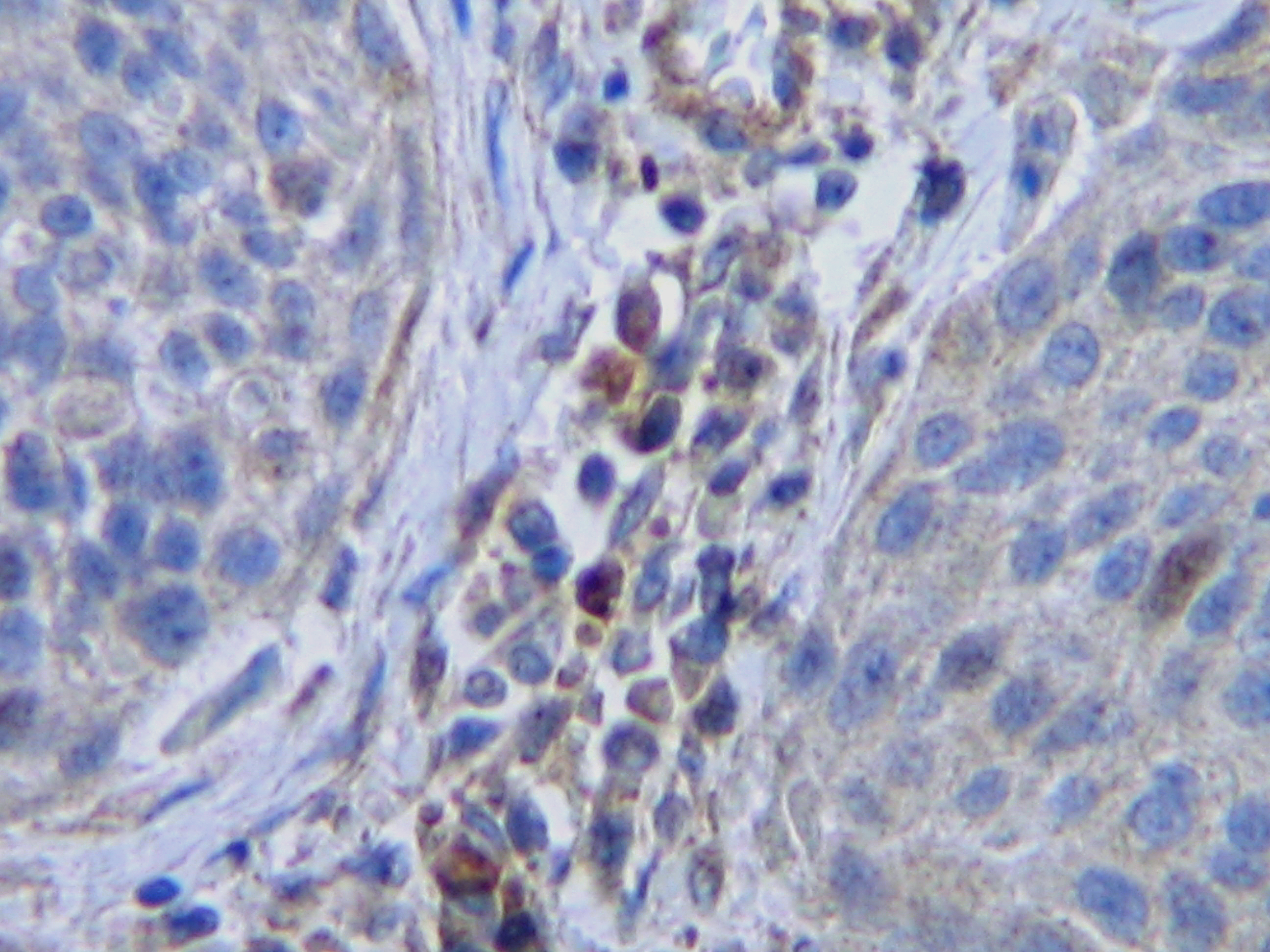
ARG66008 anti-M-CSF antibody IHC-P image
Immunohistochemistry: Formalin-fixed and paraffin-embedded sections of Human cervical squamous cell carcinoma. The recommended ARG66008 anti-M-CSF antibody concentration is 0.5 µg/ml with an overnight incubation at 4°C. An HRP-labeled polymer detection system was used with a DAB chromogen. Antigen Retrieval: Boil tissue section in Sodium Citrate buffer (pH 6.0) followed by cooling at RT for 20 min.
-
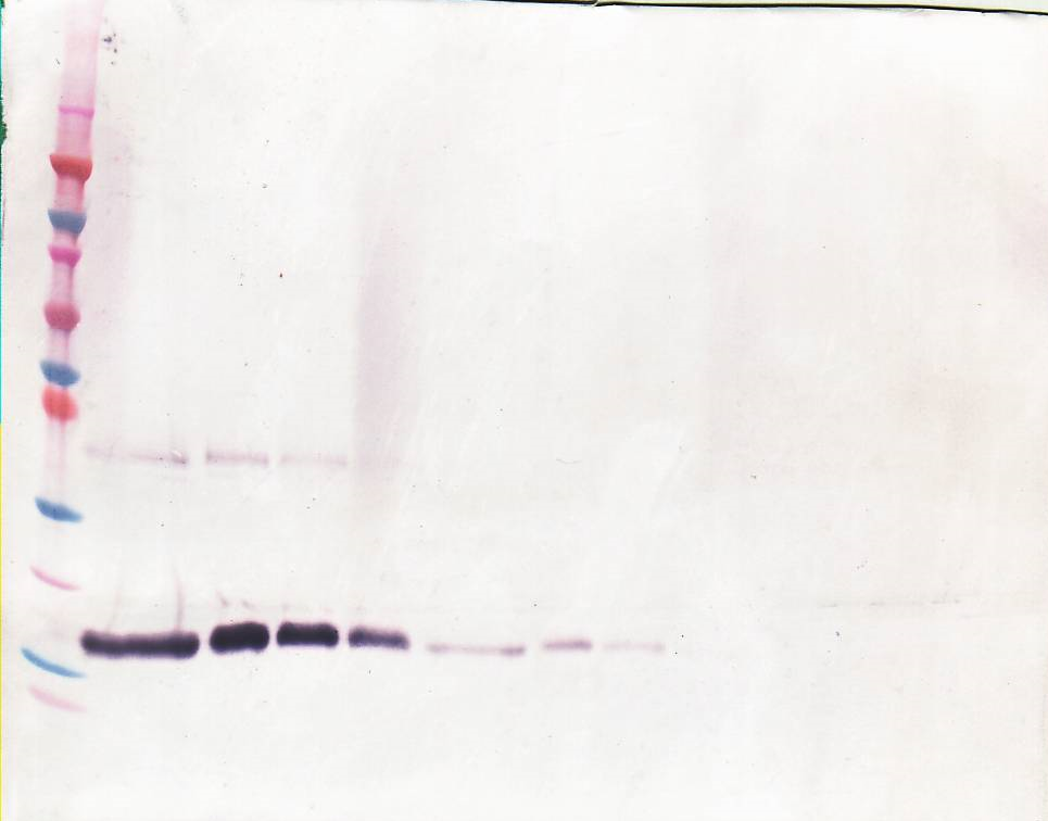
ARG66008 anti-M-CSF antibody WB image
Western blot: 250 - 0.24 ng of Human M-CSF stained with ARG66008 anti-M-CSF antibody, under non-reducing conditions.
-
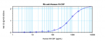
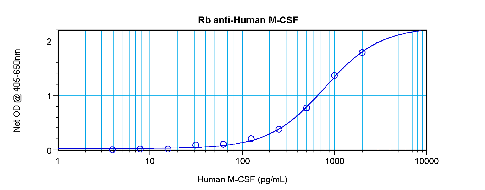
ARG66008 anti-M-CSF antibody standard curve image
Sandwich ELISA: ARG66008 anti-M-CSF antibody as a capture antibody at 0.5 - 2.0 µg/ml combined with ARG66009 anti-M-CSF antibody (Biotin) as a detection antibody. Results of a typical standard run with optical density reading at 405 - 650 nm.
-

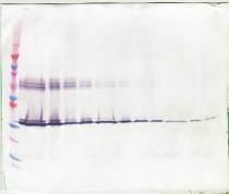
ARG66008 anti-M-CSF antibody WB image
Western blot: 250 - 0.24 ng of Human M-CSF stained with ARG66008 anti-M-CSF antibody, under reducing conditions.