ARG55381
anti-MCL1 antibody
anti-MCL1 antibody for Flow cytometry,IHC-Formalin-fixed paraffin-embedded sections,Western blot and Human,Mouse,Rat
Cancer antibody; Cell Biology and Cellular Response antibody; Cell Death antibody; Metabolism antibody

Overview
| Product Description | Rabbit Polyclonal antibody recognizes MCL1 |
|---|---|
| Tested Reactivity | Hu, Ms, Rat |
| Tested Application | FACS, IHC-P, WB |
| Host | Rabbit |
| Clonality | Polyclonal |
| Isotype | IgG |
| Target Name | MCL1 |
| Antigen Species | Human |
| Immunogen | KLH-conjugated synthetic peptide corresponding to aa. 191-226 of Human MCL1. |
| Conjugation | Un-conjugated |
| Alternate Names | Bcl-2-related protein EAT/mcl1; BCL2L3; MCL1S; Induced myeloid leukemia cell differentiation protein Mcl-1; MCL1-ES; mcl1/EAT; Bcl-2-like protein 3; EAT; Mcl-1; bcl2-L-3; Bcl2-L-3; MCL1L; TM |
Application Instructions
| Application Suggestion |
|
||||||||
|---|---|---|---|---|---|---|---|---|---|
| Application Note | * The dilutions indicate recommended starting dilutions and the optimal dilutions or concentrations should be determined by the scientist. |
Properties
| Form | Liquid |
|---|---|
| Purification | Purification with Protein G. |
| Buffer | PBS and 0.09% (W/V) Sodium azide |
| Preservative | 0.09% (W/V) Sodium azide |
| Storage Instruction | For continuous use, store undiluted antibody at 2-8°C for up to a week. For long-term storage, aliquot and store at -20°C or below. Storage in frost free freezers is not recommended. Avoid repeated freeze/thaw cycles. Suggest spin the vial prior to opening. The antibody solution should be gently mixed before use. |
| Note | For laboratory research only, not for drug, diagnostic or other use. |
Bioinformation
| Database Links | |
|---|---|
| Gene Symbol | MCL1 |
| Gene Full Name | myeloid cell leukemia 1 |
| Background | This gene encodes an anti-apoptotic protein, which is a member of the Bcl-2 family. Alternative splicing results in multiple transcript variants. The longest gene product (isoform 1) enhances cell survival by inhibiting apoptosis while the alternatively spliced shorter gene products (isoform 2 and isoform 3) promote apoptosis and are death-inducing. [provided by RefSeq, Oct 2010] |
| Function | Involved in the regulation of apoptosis versus cell survival, and in the maintenance of viability but not of proliferation. Mediates its effects by interactions with a number of other regulators of apoptosis. Isoform 1 inhibits apoptosis. Isoform 2 promotes apoptosis. [UniProt] |
| Cellular Localization | Membrane; Single-pass membrane protein. Cytoplasm. Mitochondrion. Nucleus, nucleoplasm. Note=Cytoplasmic, associated with mitochondria |
| Research Area | Cancer antibody; Cell Biology and Cellular Response antibody; Cell Death antibody; Metabolism antibody |
| Calculated MW | 37 kDa |
| PTM | Cleaved by CASP3 during apoptosis. In intact cells cleavage occurs preferentially after Asp-127, yielding a pro-apoptotic 28 kDa C-terminal fragment. Rapidly degraded in the absence of phosphorylation on Thr-163 in the PEST region. Phosphorylated on Ser-159, by GSK3, in response to IL3/interleukin-3 withdrawal. Phosphorylation at Ser-159 induces ubiquitination and proteasomal degradation, abrogating the anti-apoptotic activity. Treatment with taxol or okadaic acid induces phosphorylation on additional sites. Ubiquitinated. Ubiquitination is induced by phosphorylation at Ser-159. |
Images (4) Click the Picture to Zoom In
-
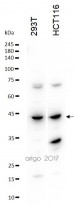
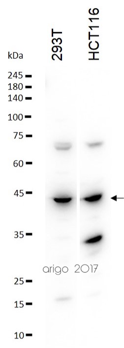
ARG55381 anti-MCL1 antibody WB image
Western blot: 30 µg of 293T and HCT116 cell lysates stained with ARG55381 anti-MCL1 antibody at 1:500 dilution.
-
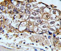
ARG55381 anti-MCL1 antibody IHC-P image
Immunohistochemistry: Formalin-fixed and paraffin-embedded Human breast carcinoma stained with ARG55381 anti-MCL1 antibody.
-
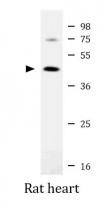
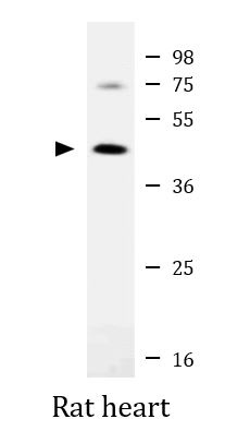
ARG55381 anti-MCL1 antibody WB image
Western blot: 20 µg of Rat heart lysate stained with ARG55381 anti-MCL1 antibody at 1:2000 dilution.
-
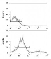
ARG55381 anti-MCL1 antibody FACS image
Flow Cytometry: ZR-75-1 cells stained with ARG55381 anti-MCL1 antibody (bottom histogram) or without primary antibody control (top histogram), followed by incubation with FITC labelled secondary antibody.


Customer's Feedback
Excellent

Review for anti-MCL1 antibody
Application:WB
Sample:293T and HCT116
Sample Loading Amount:30 µg
Primary Antibody Dilution Factor:1:500
Primary Antibody Incubation Time:overnight
Primary Antibody Incubation Temperature:4 ºC


