ARG56053
anti-MITF antibody [D5]
anti-MITF antibody [D5] for IHC-Formalin-fixed paraffin-embedded sections and Human

1
Overview
| Product Description | Mouse Monoclonal antibody [D5] recognizes MITF |
|---|---|
| Tested Reactivity | Hu |
| Species Does Not React With | Ms, Rat |
| Tested Application | IHC-P |
| Host | Mouse |
| Clonality | Monoclonal |
| Clone | D5 |
| Isotype | IgG1, kappa |
| Target Name | MITF |
| Antigen Species | Human |
| Immunogen | An N-terminus fragment of Human MITF protein |
| Conjugation | Un-conjugated |
| Alternate Names | bHLHe32; Class E basic helix-loop-helix protein 32; MI; Microphthalmia-associated transcription factor; WS2; WS2A; CMM8 |
Application Instructions
| Application Suggestion |
|
||||
|---|---|---|---|---|---|
| Application Note | IHC-P: Antigen Retrieval: Boil tissue section in 10 mM Citrate buffer (pH 6.0) for 10-20 min, followed by cooling at RT for 20 min. * The dilutions indicate recommended starting dilutions and the optimal dilutions or concentrations should be determined by the scientist. |
Properties
| Form | Liquid |
|---|---|
| Purification | Purification with Protein G. |
| Buffer | PBS (pH 7.4), 0.05% Sodium azide and 0.1 mg/ml BSA |
| Preservative | 0.05% Sodium azide |
| Stabilizer | 0.1 mg/ml BSA |
| Concentration | 0.2 mg/ml |
| Storage Instruction | For continuous use, store undiluted antibody at 2-8°C for up to a week. For long-term storage, aliquot and store at -20°C or below. Storage in frost free freezers is not recommended. Avoid repeated freeze/thaw cycles. Suggest spin the vial prior to opening. The antibody solution should be gently mixed before use. |
| Note | For laboratory research only, not for drug, diagnostic or other use. |
Bioinformation
| Database Links |
Swiss-port # O75030 Human Microphthalmia-associated transcription factor |
|---|---|
| Gene Symbol | MITF |
| Gene Full Name | microphthalmia-associated transcription factor |
| Background | This gene encodes a transcription factor that contains both basic helix-loop-helix and leucine zipper structural features. It regulates the differentiation and development of melanocytes retinal pigment epithelium and is also responsible for pigment cell-specific transcription of the melanogenesis enzyme genes. Heterozygous mutations in the this gene cause auditory-pigmentary syndromes, such as Waardenburg syndrome type 2 and Tietz syndrome. Alternatively spliced transcript variants encoding different isoforms have been identified. [provided by RefSeq, Jul 2008] |
| Function | Transcription factor that regulates the expression of genes with essential roles in cell differentiation, proliferation and survival. Binds to symmetrical DNA sequences (E-boxes) (5'-CACGTG-3') found in the promoters of target genes, such as BCL2 and tyrosinase (TYR). Plays an important role in melanocyte development by regulating the expression of tyrosinase (TYR) and tyrosinase-related protein 1 (TYRP1). Plays a critical role in the differentiation of various cell types, such as neural crest-derived melanocytes, mast cells, osteoclasts and optic cup-derived retinal pigment epithelium. [UniProt] |
| Cellular Localization | Nuclear |
| Calculated MW | 59 kDa |
| PTM | Phosphorylation at Ser-405 significantly enhances the ability to bind the tyrosinase promoter. Phosphorylated at Ser-180 and Ser-516 following KIT signaling, trigerring a short live activation: Phosphorylation at Ser-180 and Ser-516 by MAPK and RPS6KA1, respectively, activate the transcription factor activity but also promote ubiquitination and subsequent degradation by the proteasome. Ubiquitinated following phosphorylation at Ser-180, leading to subsequent degradation by the proteasome. Deubiquitinated by USP13, preventing its degradation. |
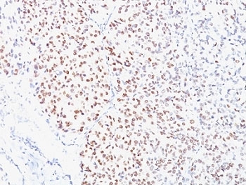
Images (1) Click the Picture to Zoom In
-
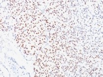
ARG56053 anti-MITF antibody [D5] IHC-P image
Immunohistochemistry: Formalin-fixed, paraffin-embedded Human melanoma stained with ARG56053 anti-MITF antibody [D5].
Customer's Feedback
Good

Review for anti-MITF antibody [D5]
Application:WB
Sample:HeLa and A549
Sample Loading Amount:30 µg
Primary Antibody Dilution Factor:1:100
Primary Antibody Incubation Time:overnight
Primary Antibody Incubation Temperature:4 ºC


