ARG55978
anti-Melanoma gp100 antibody [NKI-beteb]
anti-Melanoma gp100 antibody [NKI-beteb] for IHC-Formalin-fixed paraffin-embedded sections and Human
Overview
| Product Description | Mouse Monoclonal antibody [NKI-beteb] recognizes Melanoma gp100 |
|---|---|
| Tested Reactivity | Hu |
| Species Does Not React With | Rat |
| Tested Application | IHC-P |
| Host | Mouse |
| Clonality | Monoclonal |
| Clone | NKI-beteb |
| Isotype | IgG2b, kappa |
| Target Name | Melanoma gp100 |
| Antigen Species | Human |
| Immunogen | Membranes from a Human melanoma metastasis. |
| Conjugation | Un-conjugated |
| Alternate Names | Premelanosome protein; SILV; ME20; Melanocyte protein Pmel 17; ME20-M; Secreted melanoma-associated ME20 antigen; 95 kDa melanocyte-specific secreted glycoprotein; Silver locus protein homolog; ME20S; D12S53E; SIL; P1; Melanocyte protein PMEL; PMEL17; ME20-S; Melanoma-associated ME20 antigen; gp100; ME20M; P100; SI; P26; Melanocytes lineage-specific antigen GP100 |
Application Instructions
| Application Suggestion |
|
||||
|---|---|---|---|---|---|
| Application Note | IHC-P: Antigen Retrieval: Boil tissue section in 10 mM Citrate buffer (pH 6.0) for 10-20 min, followed by cooling at RT for 20 min. * The dilutions indicate recommended starting dilutions and the optimal dilutions or concentrations should be determined by the scientist. |
Properties
| Form | Liquid |
|---|---|
| Purification | Purification with Protein G. |
| Buffer | PBS (pH 7.4), 0.05% Sodium azide and 0.1 mg/ml BSA |
| Preservative | 0.05% Sodium azide |
| Stabilizer | 0.1 mg/ml BSA |
| Concentration | 0.2 mg/ml |
| Storage Instruction | For continuous use, store undiluted antibody at 2-8°C for up to a week. For long-term storage, aliquot and store at -20°C or below. Storage in frost free freezers is not recommended. Avoid repeated freeze/thaw cycles. Suggest spin the vial prior to opening. The antibody solution should be gently mixed before use. |
| Note | For laboratory research only, not for drug, diagnostic or other use. |
Bioinformation
| Database Links | |
|---|---|
| Gene Symbol | PMEL |
| Gene Full Name | premelanosome protein |
| Background | This gene encodes a melanocyte-specific type I transmembrane glycoprotein. The encoded protein is enriched in melanosomes, which are the melanin-producing organelles in melanocytes, and plays an essential role in the structural organization of premelanosomes. This protein is involved in generating internal matrix fibers that define the transition from Stage I to Stage II melanosomes. This protein undergoes a complex pattern of prosttranslational processing and modification that is essential to the proper functioning of the protein. A secreted form of this protein that is released by proteolytic ectodomain shedding may be used as a melanoma-specific serum marker. Alternate splicing results in multiple transcript variants. [provided by RefSeq, Jan 2011] |
| Function | Plays a central role in the biogenesis of melanosomes. Involved in the maturation of melanosomes from stage I to II. The transition from stage I melanosomes to stage II melanosomes involves an elongation of the vesicle, and the appearance within of distinct fibrillar structures. Release of the soluble form, ME20-S, could protect tumor cells from antibody mediated immunity. [UniProt] |
| Cellular Localization | Cytoplasmic |
| Calculated MW | 70 kDa |
| PTM | A small amount of P1/P100 (major form) undergoes glycosylation to yield P2/P120 (minor form). P2 is cleaved by a furin-like proprotein convertase (PC) in a pH-dependent manner in a post-Golgi, prelysosomal compartment into two disulfide-linked subunits: a large lumenal subunit, M-alpha/ME20-S, and an integral membrane subunit, M-beta. Despite cleavage, only a small fraction of M-alpha is secreted, whereas most M-alpha and M-beta remain associated with each other intracellularly. M-alpha is further processed to M-alpha N and M-alpha C. M-alpha C further undergoes processing to yield M-alpha C1 and M-alpha C3 (M-alpha C2 in the case of PMEL17-is or PMEL17-ls). Formation of intralumenal fibrils in the melanosomes requires the formation of M-alpha that becomes incorporated into the fibrils. Stage II melanosomes harbor only Golgi-modified Pmel17 fragments that are derived from M-alpha and that bear sialylated O-linked oligosaccharides. N-glycosylated. O-glycosylated; contains sialic acid. |
Images (1) Click the Picture to Zoom In
-

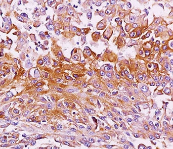
ARG55978 anti-Melanoma gp100 antibody [NKI-beteb] IHC-P image
Immunohistochemistry: Melanoma tissue stained with ARG55978 anti-Melanoma gp100 antibody [NKI-beteb].