ARG63870
anti-NOTCH3 antibody
anti-NOTCH3 antibody for Flow cytometry,IHC-Formalin-fixed paraffin-embedded sections and Human
Cell Biology and Cellular Response antibody; Developmental Biology antibody; Gene Regulation antibody; Neuroscience antibody; Signaling Transduction antibody
Overview
| Product Description | Goat Polyclonal antibody recognizes NOTCH3 |
|---|---|
| Tested Reactivity | Hu |
| Predict Reactivity | Ms, Rat |
| Tested Application | FACS, IHC-P |
| Host | Goat |
| Clonality | Polyclonal |
| Isotype | IgG |
| Target Name | NOTCH3 |
| Antigen Species | Human |
| Immunogen | C-QLGPQPEVTPKRQ |
| Conjugation | Un-conjugated |
| Alternate Names | LMNS; CADASIL; CASIL; Neurogenic locus notch homolog protein 3; IMF2; Notch 3 |
Application Instructions
| Application Suggestion |
|
||||||
|---|---|---|---|---|---|---|---|
| Application Note | IHC-P: Antigen Retrieval: Steam tissue section in Citrate buffer (pH 6.0). * The dilutions indicate recommended starting dilutions and the optimal dilutions or concentrations should be determined by the scientist. |
Properties
| Form | Liquid |
|---|---|
| Purification | Purified from goat serum by antigen affinity chromatography. |
| Buffer | Tris saline (pH 7.3), 0.02% Sodium azide and 0.5% BSA. |
| Preservative | 0.02% Sodium azide |
| Stabilizer | 0.5% BSA |
| Concentration | 0.5 mg/ml |
| Storage Instruction | For continuous use, store undiluted antibody at 2-8°C for up to a week. For long-term storage, aliquot and store at -20°C or below. Storage in frost free freezers is not recommended. Avoid repeated freeze/thaw cycles. Suggest spin the vial prior to opening. The antibody solution should be gently mixed before use. |
| Note | For laboratory research only, not for drug, diagnostic or other use. |
Bioinformation
| Database Links |
Swiss-port # Q9UM47 Human Neurogenic locus notch homolog protein 3 |
|---|---|
| Background | This gene encodes the third discovered human homologue of the Drosophilia melanogaster type I membrane protein notch. In Drosophilia, notch interaction with its cell-bound ligands (delta, serrate) establishes an intercellular signalling pathway that plays a key role in neural development. Homologues of the notch-ligands have also been identified in human, but precise interactions between these ligands and the human notch homologues remains to be determined. Mutations in NOTCH3 have been identified as the underlying cause of cerebral autosomal dominant arteriopathy with subcortical infarcts and leukoencephalopathy (CADASIL). [provided by RefSeq, Jul 2008] |
| Research Area | Cell Biology and Cellular Response antibody; Developmental Biology antibody; Gene Regulation antibody; Neuroscience antibody; Signaling Transduction antibody |
| Calculated MW | 244 kDa |
| PTM | Synthesized in the endoplasmic reticulum as an inactive form which is proteolytically cleaved by a furin-like convertase in the trans-Golgi network before it reaches the plasma membrane to yield an active, ligand-accessible form. Cleavage results in a C-terminal fragment N(TM) and a N-terminal fragment N(EC). Following ligand binding, it is cleaved by TNF-alpha converting enzyme (TACE) to yield a membrane-associated intermediate fragment called notch extracellular truncation (NEXT). This fragment is then cleaved by presenilin dependent gamma-secretase to release a notch-derived peptide containing the intracellular domain (NICD) from the membrane (By similarity). Phosphorylated. Hydroxylated by HIF1AN. |
Images (3) Click the Picture to Zoom In
-
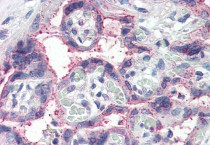
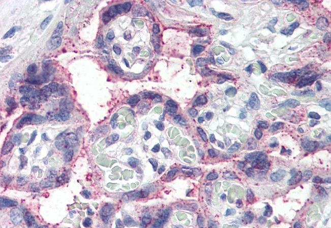
ARG63870 anti-Notch 3 antibody IHC-P image
Immunohistochemistry: Paraffin-embedded Human placenta tissue. Antigen Retrieval: Steam tissue section in Citrate buffer (pH 6.0). The tissue section was stained with ARG63870 anti-Notch 3 antibody at 5 µg/ml dilution followed by AP-staining.
-
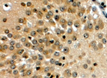
ARG63870 anti-Notch 3 antibody IHC-P image
Immunohistochemistry: Paraffin embedded Human Hippocampus. (Steamed antigen retrieval with citrate buffer pH 6) stained with ARG63870 anti-Notch 3 antibody at 4 µg/ml dilution followed by HRP-staining.
-
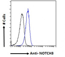
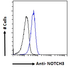
ARG63870 anti-Notch 3 antibody FACS image
Flow Cytometry: Paraformaldehyde-fixed HeLa cells permeabilized with 0.5% Triton. Cells were stained with ARG63870 anti-Notch 3 antibody (blue line) at 10 µg/ml dilution for 1 hour, followed by incubation with Alexa FluorR 488 labelled secondary antibody. IgG control: Unimmunized goat IgG (black line).