ARG42975
anti-NOVA2 antibody
anti-NOVA2 antibody for Flow cytometry,IHC-Formalin-fixed paraffin-embedded sections,Western blot and Human,Mouse,Rat
Overview
| Product Description | Rabbit Polyclonal antibody recognizes NOVA2 |
|---|---|
| Tested Reactivity | Hu, Ms, Rat |
| Tested Application | FACS, IHC-P, WB |
| Host | Rabbit |
| Clonality | Polyclonal |
| Isotype | IgG |
| Target Name | NOVA2 |
| Antigen Species | Human |
| Immunogen | Recombinant protein corresponding to M1-Q205 of Human NOVA2. |
| Conjugation | Un-conjugated |
| Alternate Names | NOVA3; RNA-binding protein Nova-2; Astrocytic NOVA1-like RNA-binding protein; ANOVA; Neuro-oncological ventral antigen 2 |
Application Instructions
| Application Suggestion |
|
||||||||
|---|---|---|---|---|---|---|---|---|---|
| Application Note | IHC-P: Antigen Retrieval: Heat mediation was performed in EDTA buffer (pH 8.0). * The dilutions indicate recommended starting dilutions and the optimal dilutions or concentrations should be determined by the scientist. |
||||||||
| Observed Size | 49, 72 kDa |
Properties
| Form | Liquid |
|---|---|
| Purification | Affinity purification with immunogen. |
| Buffer | 0.2% Na2HPO4, 0.9% NaCl, 0.05% Sodium azide and 4% Trehalose. |
| Preservative | 0.05% Sodium azide |
| Stabilizer | 4% Trehalose |
| Concentration | 0.5 mg/ml |
| Storage Instruction | For continuous use, store undiluted antibody at 2-8°C for up to a week. For long-term storage, aliquot and store at -20°C or below. Storage in frost free freezers is not recommended. Avoid repeated freeze/thaw cycles. Suggest spin the vial prior to opening. The antibody solution should be gently mixed before use. |
| Note | For laboratory research only, not for drug, diagnostic or other use. |
Bioinformation
| Database Links | |
|---|---|
| Gene Symbol | NOVA2 |
| Gene Full Name | neuro-oncological ventral antigen 2 |
| Function | May regulate RNA splicing or metabolism in a specific subset of developing neurons (By similarity). Binds single strand RNA. [UniProt] |
| Cellular Localization | Nucleus. [UniProt] |
| Calculated MW | 49 kDa |
Images (7) Click the Picture to Zoom In
-
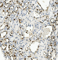
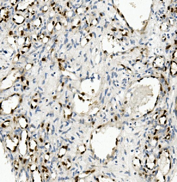
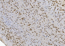
ARG42975 anti-NOVA2 antibody IHC-P image
Immunohistochemistry: Paraffin-embedded Human appendicitis tissue. Antigen Retrieval: Heat mediation was performed in EDTA buffer (pH 8.0). The tissue section was blocked with 10% goat serum. The tissue section was then stained with ARG42975 anti-NOVA2 antibody at 1 µg/ml dilution, overnight at 4°C.
-
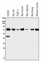
ARG42975 anti-NOVA2 antibody WB image
Western blot: 50 µg of sample under reducing conditions. U2OS, HL-60, THP-1, Rat brain, Rat spleen, Rat lung and Mouse brain lysates stained with ARG42975 anti-NOVA2 antibody at 0.5 µg/ml dilution, overnight at 4°C.
-
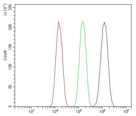
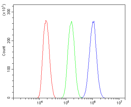
ARG42975 anti-NOVA2 antibody FACS image
Flow Cytometry: A549 cells were blocked with 10% normal goat serum and then stained with ARG42975 anti-NOVA2 antibody (blue) at 1 µg/10^6 cells for 30 min at 20°C, followed by incubation with DyLight®488 labelled secondary antibody. Isotype control antibody (green) was Rabbit IgG (1 µg/10^6 cells) used under the same conditions. Unlabelled sample (red) was also used as a control.
-
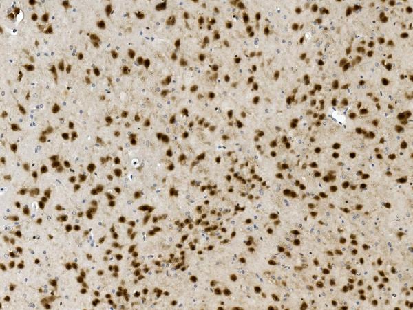
ARG42975 anti-NOVA2 antibody IHC-P image
Immunohistochemistry: Paraffin-embedded Mouse brain tissue. Antigen Retrieval: Heat mediation was performed in EDTA buffer (pH 8.0). The tissue section was blocked with 10% goat serum. The tissue section was then stained with ARG42975 anti-NOVA2 antibody at 1 µg/ml dilution, overnight at 4°C.
-
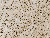
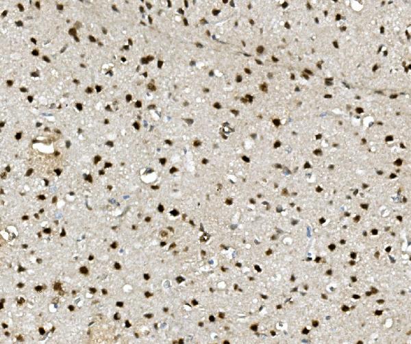
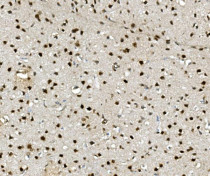
ARG42975 anti-NOVA2 antibody IHC-P image
Immunohistochemistry: Paraffin-embedded Rat liver tissue. Antigen Retrieval: Heat mediation was performed in EDTA buffer (pH 8.0). The tissue section was blocked with 10% goat serum. The tissue section was then stained with ARG42975 anti-NOVA2 antibody at 1 µg/ml dilution, overnight at 4°C.
-
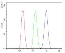
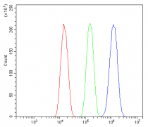
ARG42975 anti-NOVA2 antibody FACS image
Flow Cytometry: Hepa 1-6 cells were blocked with 10% normal goat serum and then stained with ARG42975 anti-NOVA2 antibody (blue) at 1 µg/10^6 cells for 30 min at 20°C, followed by incubation with DyLight®488 labelled secondary antibody. Isotype control antibody (green) was Rabbit IgG (1 µg/10^6 cells) used under the same conditions. Unlabelled sample (red) was also used as a control.