ARG10800
anti-PDE4A antibody
anti-PDE4A antibody for Confocal microscopy (Confocal),Dot blot,ELISA,ICC/IF,IHC-Formalin-fixed paraffin-embedded sections,Immunoprecipitation,Western blot and Human,Mouse,Rat
Overview
| Product Description | Rabbit Polyclonal antibody recognizes PDE4A |
|---|---|
| Tested Reactivity | Hu, Ms, Rat |
| Tested Application | Confocal, Dot, ELISA, ICC/IF, IHC-P, IP, WB |
| Host | Rabbit |
| Clonality | Polyclonal |
| Isotype | IgG |
| Target Name | PDE4A |
| Antigen Species | Human |
| Immunogen | Synthetic cyclic peptide around the C-terminus of PDE4A. Common to all PDE4A proteins. |
| Conjugation | Un-conjugated |
| Alternate Names | DPDE2; cAMP-specific 3',5'-cyclic phosphodiesterase 4A; PDE4; PDE46; EC 3.1.4.53 |
Application Instructions
| Application Suggestion |
|
||||||||||||||||
|---|---|---|---|---|---|---|---|---|---|---|---|---|---|---|---|---|---|
| Application Note | * The dilutions indicate recommended starting dilutions and the optimal dilutions or concentrations should be determined by the scientist. |
Properties
| Form | Liquid |
|---|---|
| Purification | Affinity purified. |
| Buffer | Tris-Glycine Buffer (pH 7.4 - 7.8), Hepes, 0.02% Sodium azide, 30% Glycerol and 0.5% BSA. |
| Preservative | 0.02% Sodium azide |
| Stabilizer | 30% Glycerol and 0.5% BSA |
| Concentration | 0.5 mg/ml |
| Storage Instruction | For continuous use, store undiluted antibody at 2-8°C for up to a week. For long-term storage, aliquot and store at -20°C. Storage in frost free freezers is not recommended. Avoid repeated freeze/thaw cycles. Suggest spin the vial prior to opening. The antibody solution should be gently mixed before use. |
| Note | For laboratory research only, not for drug, diagnostic or other use. |
Bioinformation
| Database Links | |
|---|---|
| Gene Symbol | PDE4A |
| Gene Full Name | phosphodiesterase 4A, cAMP-specific |
| Background | The protein encoded by this gene belongs to the cyclic nucleotide phosphodiesterase (PDE) family, and PDE4 subfamily. This PDE hydrolyzes the second messenger, cAMP, which is a regulator and mediator of a number of cellular responses to extracellular signals. Thus, by regulating the cellular concentration of cAMP, this protein plays a key role in many important physiological processes. Alternatively spliced transcript variants encoding different isoforms have been described for this gene.[provided by RefSeq, Jul 2011] |
| Function | Hydrolyzes the second messenger cAMP, which is a key regulator of many important physiological processes. [UniProt] |
| Calculated MW | 98 kDa |
| PTM | Phosphorylation by MAPKAPK2 its activation through PKA phosphorylation (By similarity). Phosphorylated at Ser-686 and Ser-688 when expressed in S.frugiperda cells. Isoform 2 and isoform 7 are activated by phosphorylation at Ser-119 and Ser-123 respectively by PKA. Proteolytically cleaved by caspase-3. |
Images (6) Click the Picture to Zoom In
-

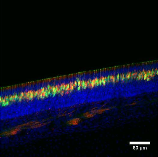
ARG10800 anti-PDE4A antibody IHC image
Immunohistochemistry: Thick section of Mouse nose tissue stained with ARG10800 anti-PDE4A antibody (red). Green: Olfactory sensory neurons. Blue: Nuclei.
-
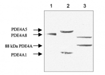
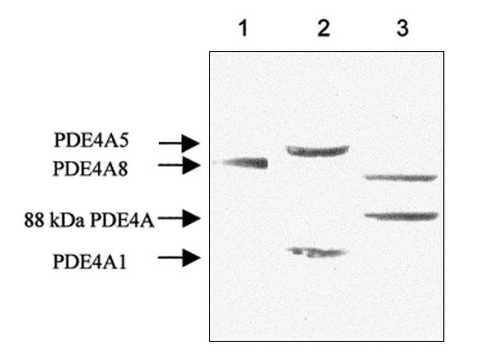
ARG10800 anti-PDE4A antibody WB image
Western blot: 1) Recombinant PDE4A8, 2) Recombinant PDE4A5 & PDE4A1, and 3) Rat testis lysates stained with ARG10800 anti-PDE4A antibody at 1:500 dilution.
-

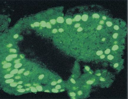
ARG10800 anti-PDE4A antibody Confocal image
Scanning laser confocal microscopy: Fixed section of Rat seminefrous tubule stained with ARG10800 anti-PDE4A antibody.
-
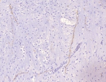
ARG10800 anti-PDE4A antibody IHC-P image
Immunohistochemistry: Paraffin-embedded Human ovarian medulla tissue stained with ARG10800 anti-PDE4A antibody.
-
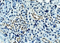
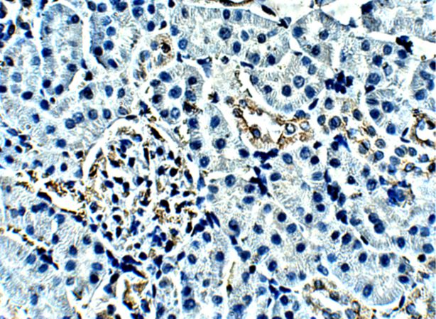
ARG10800 anti-PDE4A antibody IHC-P image
Immunohistochemistry: Paraffin-embedded Rat kidney tissue stained with ARG10800 anti-PDE4A antibody at 1:100 dilution.
-
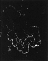
ARG10800 anti-PDE4A antibody IHC-Fr image
Immunohistochemistry: Frozen section of Rat seminiferous tubule stained with ARG10800 anti-PDE4A antibody.