ARG55525
anti-PKC iota antibody
anti-PKC iota antibody for IHC-Formalin-fixed paraffin-embedded sections,Western blot and Human
Signaling Transduction antibody
Overview
| Product Description | Rabbit Polyclonal antibody recognizes PKC iota |
|---|---|
| Tested Reactivity | Hu |
| Tested Application | IHC-P, WB |
| Host | Rabbit |
| Clonality | Polyclonal |
| Isotype | IgG |
| Target Name | PKC iota |
| Antigen Species | Human |
| Immunogen | KLH-conjugated synthetic peptide corresponding to aa. 176-207 (N-terminus) of Human PKC iota. |
| Conjugation | Un-conjugated |
| Alternate Names | PRKC-lambda/iota; EC 2.7.11.13; Protein kinase C iota type; nPKC-iota; PKCI; Atypical protein kinase C-lambda/iota; DXS1179E; aPKC-lambda/iota |
Application Instructions
| Application Suggestion |
|
||||||
|---|---|---|---|---|---|---|---|
| Application Note | * The dilutions indicate recommended starting dilutions and the optimal dilutions or concentrations should be determined by the scientist. |
Properties
| Form | Liquid |
|---|---|
| Purification | Purification with Protein G. |
| Buffer | PBS and 0.09% (W/V) Sodium azide |
| Preservative | 0.09% (W/V) Sodium azide |
| Storage Instruction | For continuous use, store undiluted antibody at 2-8°C for up to a week. For long-term storage, aliquot and store at -20°C or below. Storage in frost free freezers is not recommended. Avoid repeated freeze/thaw cycles. Suggest spin the vial prior to opening. The antibody solution should be gently mixed before use. |
| Note | For laboratory research only, not for drug, diagnostic or other use. |
Bioinformation
| Database Links | |
|---|---|
| Gene Symbol | PRKCI |
| Gene Full Name | protein kinase C, iota |
| Background | This gene encodes a member of the protein kinase C (PKC) family of serine/threonine protein kinases. The PKC family comprises at least eight members, which are differentially expressed and are involved in a wide variety of cellular processes. This protein kinase is calcium-independent and phospholipid-dependent. It is not activated by phorbolesters or diacylglycerol. This kinase can be recruited to vesicle tubular clusters (VTCs) by direct interaction with the small GTPase RAB2, where this kinase phosphorylates glyceraldehyde-3-phosphate dehydrogenase (GAPD/GAPDH) and plays a role in microtubule dynamics in the early secretory pathway. This kinase is found to be necessary for BCL-ABL-mediated resistance to drug-induced apoptosis and therefore protects leukemia cells against drug-induced apoptosis. There is a single exon pseudogene mapped on chromosome X. [provided by RefSeq, Jul 2008] |
| Function | Calcium- and diacylglycerol-independent serine/ threonine-protein kinase that plays a general protective role against apoptotic stimuli, is involved in NF-kappa-B activation, cell survival, differentiation and polarity, and contributes to the regulation of microtubule dynamics in the early secretory pathway. Is necessary for BCR-ABL oncogene-mediated resistance to apoptotic drug in leukemia cells, protecting leukemia cells against drug-induced apoptosis. In cultured neurons, prevents amyloid beta protein-induced apoptosis by interrupting cell death process at a very early step. In glioblastoma cells, may function downstream of phosphatidylinositol 3-kinase (PI(3)K) and PDPK1 in the promotion of cell survival by phosphorylating and inhibiting the pro-apoptotic factor BAD. Can form a protein complex in non-small cell lung cancer (NSCLC) cells with PARD6A and ECT2 and regulate ECT2 oncogenic activity by phosphorylation, which in turn promotes transformed growth and invasion. In response to nerve growth factor (NGF), acts downstream of SRC to phosphorylate and activate IRAK1, allowing the subsequent activation of NF-kappa-B and neuronal cell survival. Functions in the organization of the apical domain in epithelial cells by phosphorylating EZR. This step is crucial for activation and normal distribution of EZR at the early stages of intestinal epithelial cell differentiation. Forms a protein complex with LLGL1 and PARD6B independently of PARD3 to regulate epithelial cell polarity. Plays a role in microtubule dynamics in the early secretory pathway through interaction with RAB2A and GAPDH and recruitment to vesicular tubular clusters (VTCs). In human coronary artery endothelial cells (HCAEC), is activated by saturated fatty acids and mediates lipid-induced apoptosis. [UniProt] |
| Research Area | Signaling Transduction antibody |
| Calculated MW | 68 kDa |
| PTM | Phosphorylation at Thr-412 in the activation loop is not mandatory for activation (By similarity). Upon neuronal growth factor (NGF) stimulation, phosphorylated by SRC at Tyr-265, Tyr-280 and Tyr-334. Phosphorylation at Tyr-265 facilitates binding to KPNB1/importin-beta regulating entry of PRKCI into the nucleus. Phosphorylation on Tyr-334 is important for NF-kappa-B stimulation. |
Images (2) Click the Picture to Zoom In
-
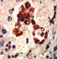
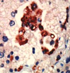
ARG55525 anti-PKC iota antibody IHC-P image
Immunohistochemistry: Formalin-fixed and paraffin-embedded Human cancer tissue stained with ARG55525 anti-PKC iota antibody.
-
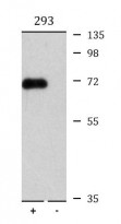
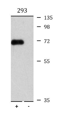
ARG55525 anti-PKC iota antibody WB image
Western blot: 2 µg of 293 cell lysate either 1) transiently transfected with PKC iota gene or 2) nontransfected. The blots were stained with ARG55525 anti-PKC iota antibody.