ARG63589
anti-Pleckstrin antibody
anti-Pleckstrin antibody for Flow cytometry,ICC/IF,IHC-Formalin-fixed paraffin-embedded sections,Western blot and Human
Cell Biology and Cellular Response antibody; Signaling Transduction antibody
Overview
| Product Description | Goat Polyclonal antibody recognizes Pleckstrin |
|---|---|
| Tested Reactivity | Hu |
| Tested Application | FACS, ICC/IF, IHC-P, WB |
| Host | Goat |
| Clonality | Polyclonal |
| Isotype | IgG |
| Target Name | Pleckstrin |
| Antigen Species | Human |
| Immunogen | C-EWIKAIQMASRTGK |
| Conjugation | Un-conjugated |
| Alternate Names | P47; p47; Pleckstrin; Platelet 47 kDa protein |
Application Instructions
| Application Suggestion |
|
||||||||||
|---|---|---|---|---|---|---|---|---|---|---|---|
| Application Note | WB: Recommend incubate at RT for 1h. IHC-P: Antigen Retrieval: Steam tissue section in Citrate buffer (pH 6.0). * The dilutions indicate recommended starting dilutions and the optimal dilutions or concentrations should be determined by the scientist. |
Properties
| Form | Liquid |
|---|---|
| Purification | Purified from goat serum by antigen affinity chromatography. |
| Buffer | Tris saline (pH 7.3), 0.02% Sodium azide and 0.5% BSA. |
| Preservative | 0.02% Sodium azide |
| Stabilizer | 0.5% BSA |
| Concentration | 0.5 mg/ml |
| Storage Instruction | For continuous use, store undiluted antibody at 2-8°C for up to a week. For long-term storage, aliquot and store at -20°C or below. Storage in frost free freezers is not recommended. Avoid repeated freeze/thaw cycles. Suggest spin the vial prior to opening. The antibody solution should be gently mixed before use. |
| Note | For laboratory research only, not for drug, diagnostic or other use. |
Bioinformation
| Database Links | |
|---|---|
| Gene Symbol | PLEK |
| Gene Full Name | pleckstrin |
| Function | Major protein kinase C substrate of platelets. [UniProt] |
| Research Area | Cell Biology and Cellular Response antibody; Signaling Transduction antibody |
| Calculated MW | 40 kDa |
Images (6) Click the Picture to Zoom In
-
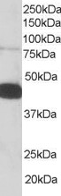
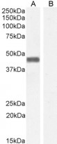
ARG63589 anti-Pleckstrin antibody WB image
Western Blot: Human PBMC lysate (RIPA buffer, 35 µg total protein per lane) stained with ARG63589 anti-Pleckstrin antibody at 0.05 µg/ml dilution.
-
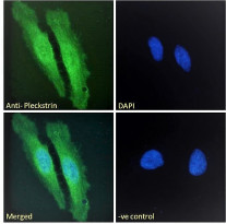
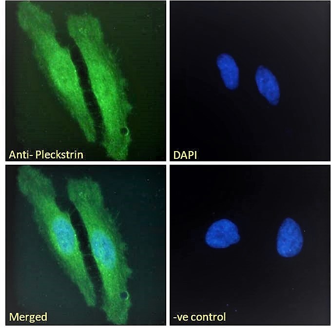
ARG63589 anti-Pleckstrin antibody ICC/IF image
Immunofluorescence: Paraformaldehyde fixed HeLa cells permeabilized with 0.15% Triton. Cells were stained with ARG63589 anti-Pleckstrin antibody (green) at 10 µg/ml dilution for 1 hour. DAPI (blue) for nuclear staining. Negative control: Unimmunized goat IgG (green) at 10 µg/ml dilution.
-

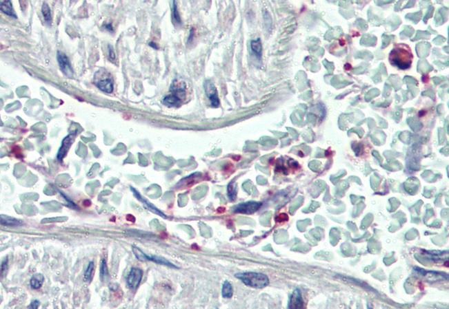
ARG63589 anti-Pleckstrin antibody IHC-P image
Immunohistochemistry: Paraffin-embedded Human vessel (platelets) tissue. Antigen Retrieval: Steam tissue section in Citrate buffer (pH 6.0). The tissue section was stained with ARG63589 anti-Pleckstrin antibody at 3.75 µg/ml dilution followed by AP-staining.
-
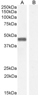
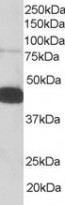
ARG63589 anti-Pleckstrin antibody WB image
Western blot: 35 µg of Human parathyroid (A) and HepG2 (B, negative control) lysates (in RIPA buffer) stained with ARG63589 anti-Pleckstrin antibody at 0.01 µg/ml dilution and incubated at RT for 1 hour.
-

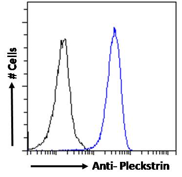
ARG63589 anti-Pleckstrin antibody FACS image
Flow Cytometry: Paraformaldehyde-fixed HeLa cells permeabilized with 0.5% Triton. Cells were stained with ARG63589 anti-Pleckstrin antibody (blue line) at 10 µg/ml dilution for 1 hour, followed by incubation with Alexa FluorR 488 labelled secondary antibody. IgG control: Unimmunized goat IgG (black line).
-
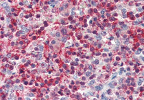
ARG63589 anti-Pleckstrin antibody IHC-P image
Immunohistochemistry: Paraffin-embedded Human spleen tissue. Antigen Retrieval: Steam tissue section in Citrate buffer (pH 6.0). The tissue section was stained with ARG63589 anti-Pleckstrin antibody at 3.75 µg/ml dilution followed by AP-staining.