ARG41418
anti-Protein C antibody
anti-Protein C antibody for Flow cytometry,IHC-Formalin-fixed paraffin-embedded sections and Human
Overview
| Product Description | Rabbit Polyclonal antibody recognizes Protein C |
|---|---|
| Tested Reactivity | Hu |
| Tested Application | FACS, IHC-P |
| Host | Rabbit |
| Clonality | Polyclonal |
| Isotype | IgG |
| Target Name | Protein C |
| Antigen Species | Human |
| Immunogen | Synthetic peptide corresponding to aa. 446-461 of Human Protein C. (HGHIRDKEAPQKSWAP) |
| Conjugation | Un-conjugated |
| Alternate Names | EC 3.4.21.69; Blood coagulation factor XIV; PC; THPH3; Vitamin K-dependent protein C; THPH4; APC; Autoprothrombin IIA; PROC1; Anticoagulant protein C |
Application Instructions
| Application Suggestion |
|
||||||
|---|---|---|---|---|---|---|---|
| Application Note | IHC-P: Antigen Retrieval: Heat mediation was performed in Citrate buffer (pH 6.0, epitope retrieval solution) for 20 min. * The dilutions indicate recommended starting dilutions and the optimal dilutions or concentrations should be determined by the scientist. |
Properties
| Form | Liquid |
|---|---|
| Purification | Affinity purification with immunogen. |
| Buffer | 0.2% Na2HPO4, 0.9% NaCl, 0.05% Thimerosal, 0.05% Sodium azide and 5% BSA. |
| Preservative | 0.05% Thimerosal and 0.05% Sodium azide |
| Stabilizer | 5% BSA |
| Concentration | 0.5 mg/ml |
| Storage Instruction | For continuous use, store undiluted antibody at 2-8°C for up to a week. For long-term storage, aliquot and store at -20°C or below. Storage in frost free freezers is not recommended. Avoid repeated freeze/thaw cycles. Suggest spin the vial prior to opening. The antibody solution should be gently mixed before use. |
| Note | For laboratory research only, not for drug, diagnostic or other use. |
Bioinformation
| Database Links | |
|---|---|
| Gene Symbol | PROC |
| Gene Full Name | protein C (inactivator of coagulation factors Va and VIIIa) |
| Background | This gene encodes a vitamin K-dependent plasma glycoprotein. The encoded protein is cleaved to its activated form by the thrombin-thrombomodulin complex. This activated form contains a serine protease domain and functions in degradation of the activated forms of coagulation factors V and VIII. Mutations in this gene have been associated with thrombophilia due to protein C deficiency, neonatal purpura fulminans, and recurrent venous thrombosis.[provided by RefSeq, Dec 2009] |
| Function | Protein C is a vitamin K-dependent serine protease that regulates blood coagulation by inactivating factors Va and VIIIa in the presence of calcium ions and phospholipids. [UniProt] |
| Cellular Localization | Secreted. Golgi apparatus. Endoplasmic reticulum. [UniProt] |
| Calculated MW | 52 kDa |
| PTM | The vitamin K-dependent, enzymatic carboxylation of some Glu residues allows the modified protein to bind calcium. N- and O-glycosylated. Partial (70%) N-glycosylation of Asn-371 with an atypical N-X-C site produces a higher molecular weight form referred to as alpha. The lower molecular weight form, not N-glycosylated at Asn-371, is beta. O-glycosylated with core 1 or possibly core 8 glycans. The iron and 2-oxoglutarate dependent 3-hydroxylation of aspartate and asparagine is (R) stereospecific within EGF domains. May be phosphorylated on a Ser or Thr in a region (AA 25-30) of the propeptide. [UniProt] |
Images (2) Click the Picture to Zoom In
-
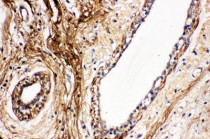
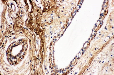
ARG41418 anti-Protein C antibody IHC-P image
Immunohistochemistry: Paraffin-embedded Human mammary cancer tissue. Antigen Retrieval: Heat mediation was performed in Citrate buffer (pH 6.0, epitope retrieval solution) for 20 min. The tissue section was blocked with 10% goat serum. The tissue section was then stained with ARG41418 anti-Protein C antibody at 1 µg/ml dilution, overnight at 4°C.
-
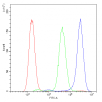
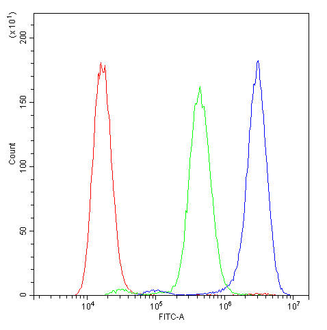
ARG41418 anti-Protein C antibody FACS image
Flow Cytometry: A549 cells were blocked with 10% normal goat serum and then stained with ARG41418 anti-Protein C antibody (blue) at 1 µg/10^6 cells for 30 min at 20°C, followed by incubation with DyLight®488 labelled secondary antibody. Isotype control antibody (green) was Rabbit IgG (1 µg/10^6 cells) used under the same conditions. Unlabelled sample (red) was also used as a control.