ARG41218
anti-RAB3D antibody
anti-RAB3D antibody for Flow cytometry,IHC-Formalin-fixed paraffin-embedded sections,Western blot and Human
Overview
| Product Description | Rabbit Polyclonal antibody recognizes RAB3D |
|---|---|
| Tested Reactivity | Hu |
| Tested Application | FACS, IHC-P, WB |
| Host | Rabbit |
| Clonality | Polyclonal |
| Isotype | IgG |
| Target Name | RAB3D |
| Antigen Species | Human |
| Immunogen | KLH-conjugated synthetic peptide corresponding to aa. 185-212 of Human RAB3D. |
| Conjugation | Un-conjugated |
| Alternate Names | RAB16; GOV; Ras-related protein Rab-3D; RAD3D; D2-2 |
Application Instructions
| Application Suggestion |
|
||||||||
|---|---|---|---|---|---|---|---|---|---|
| Application Note | * The dilutions indicate recommended starting dilutions and the optimal dilutions or concentrations should be determined by the scientist. | ||||||||
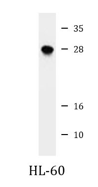
| Positive Control | HL-60 | ||||||||
| Observed Size | 28 kDa |
Properties
| Form | Liquid |
|---|---|
| Purification | Purification with Protein A and immunogen peptide. |
| Buffer | PBS and 0.09% (W/V) Sodium azide. |
| Preservative | 0.09% (W/V) Sodium azide. |
| Storage Instruction | For continuous use, store undiluted antibody at 2-8°C for up to a week. For long-term storage, aliquot and store at -20°C or below. Storage in frost free freezers is not recommended. Avoid repeated freeze/thaw cycles. Suggest spin the vial prior to opening. The antibody solution should be gently mixed before use. |
| Note | For laboratory research only, not for drug, diagnostic or other use. |
Bioinformation
| Database Links | |
|---|---|
| Gene Symbol | RAB3D |
| Gene Full Name | RAB3D, member RAS oncogene family |
| Function | Protein transport. Probably involved in regulated exocytosis (By similarity). [UniProt] |
| Cellular Localization | Cell membrane; Lipid-anchor; Cytoplasmic side. [UniProt] |
| Calculated MW | 24 kDa |
Images (3) Click the Picture to Zoom In
-

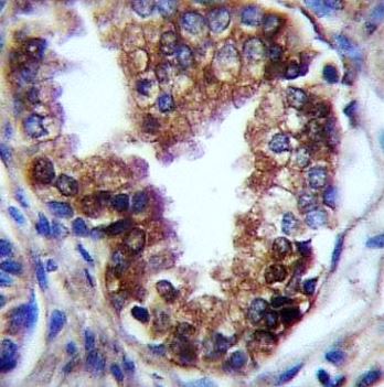
ARG41218 anti-RAB3D antibody IHC-P image
Immunohistochemistry: Formalin-fixed and paraffin-embedded Human prostate carcinoma tissue stained with ARG41218 anti-RAB3D antibody.
-

ARG41218 anti-RAB3D antibody WB image
Western blot: 35 ug of HL-60 cell lysate stained with ARG41218 anti-RAB3D antibody.
-
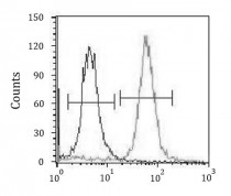
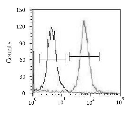
ARG41218 anti-RAB3D antibody FACS image
Flow Cytometry: K562 cells stained with ARG41218 anti-RAB3D antibody (right histogram) or without primary antibody as control (left histogram), followed by incubation with FITC labelled secondary antibody.