ARG42511
anti-RAB5B antibody
anti-RAB5B antibody for ICC/IF,IHC-Formalin-fixed paraffin-embedded sections,IHC-Frozen sections,Western blot and Dog,Human,Monkey,Mouse,Rat
Overview
| Product Description | Goat Polyclonal antibody recognizes RAB5B |
|---|---|
| Tested Reactivity | Hu, Ms, Rat, Dog, Mk |
| Tested Application | ICC/IF, IHC-Fr, IHC-P, WB |
| Host | Goat |
| Clonality | Polyclonal |
| Isotype | IgG |
| Target Name | RAB5B |
| Antigen Species | Mouse |
| Immunogen | Purified recombinant peptide within aa. 115 to the C-terminus of Mouse RAB5B. |
| Conjugation | Un-conjugated |
| Alternate Names | Ras-related protein Rab-5B |
Application Instructions
| Application Suggestion |
|
||||||||||
|---|---|---|---|---|---|---|---|---|---|---|---|
| Application Note | * The dilutions indicate recommended starting dilutions and the optimal dilutions or concentrations should be determined by the scientist. | ||||||||||
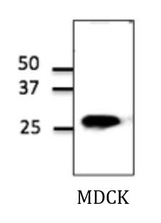
| Positive Control | MDCK | ||||||||||
| Observed Size | ~ 26 kDa |
Properties
| Form | Liquid |
|---|---|
| Purification | Affinity purification with immunogen. |
| Buffer | PBS, 0.05% Sodium azide and 20% Glycerol. |
| Preservative | 0.05% Sodium azide |
| Stabilizer | 20% Glycerol |
| Concentration | 4 mg/ml |
| Storage Instruction | For continuous use, store undiluted antibody at 2-8°C for up to a week. For long-term storage, aliquot and store at -20°C. Storage in frost free freezers is not recommended. Avoid repeated freeze/thaw cycles. Suggest spin the vial prior to opening. The antibody solution should be gently mixed before use. |
| Note | For laboratory research only, not for drug, diagnostic or other use. |
Bioinformation
| Database Links | |
|---|---|
| Gene Symbol | RAB5B |
| Gene Full Name | RAB5B, member RAS oncogene family |
| Function | Protein transport. Probably involved in vesicular traffic (By similarity). [UniProt] |
| Cellular Localization | Cell membrane; Lipid-anchor; Cytoplasmic side. Early endosome membrane; Lipid-anchor. Melanosome. Note=Enriched in stage I melanosomes. [UniProt] |
| Calculated MW | 24 kDa |
Images (2) Click the Picture to Zoom In
-

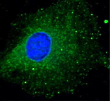
ARG42511 anti-RAB5B antibody ICC/IF image
Immunofluorescence: B6-RPE07 cells were stained with ARG42511 anti-RAB5B antibody (green) at 1:100 dilution. Nuclear staining (blue).
-

ARG42511 anti-RAB5B antibody WB image
Western blot: MDCK cell lysate stained with ARG42511 anti-RAB5B antibody.