ARG58289
anti-RPL4 antibody
anti-RPL4 antibody for Flow cytometry,ICC/IF,Western blot and Human
Overview
| Product Description | Rabbit Polyclonal antibody recognizes RPL4 |
|---|---|
| Tested Reactivity | Hu |
| Tested Application | FACS, ICC/IF, WB |
| Host | Rabbit |
| Clonality | Polyclonal |
| Isotype | IgG |
| Target Name | RPL4 |
| Antigen Species | Human |
| Immunogen | KLH-conjugated synthetic peptide corresponding to aa. 119-149 of Human RPL4. |
| Conjugation | Un-conjugated |
| Alternate Names | L4; 60S ribosomal protein L1; 60S ribosomal protein L4 |
Application Instructions
| Application Suggestion |
|
||||||||
|---|---|---|---|---|---|---|---|---|---|
| Application Note | * The dilutions indicate recommended starting dilutions and the optimal dilutions or concentrations should be determined by the scientist. | ||||||||
| Positive Control | HeLa |
Properties
| Form | Liquid |
|---|---|
| Purification | Purification with Protein A and immunogen peptide. |
| Buffer | PBS and 0.09% (W/V) Sodium azide. |
| Preservative | 0.09% (W/V) Sodium azide |
| Storage Instruction | For continuous use, store undiluted antibody at 2-8°C for up to a week. For long-term storage, aliquot and store at -20°C or below. Storage in frost free freezers is not recommended. Avoid repeated freeze/thaw cycles. Suggest spin the vial prior to opening. The antibody solution should be gently mixed before use. |
| Note | For laboratory research only, not for drug, diagnostic or other use. |
Bioinformation
| Database Links | |
|---|---|
| Gene Symbol | RPL4 |
| Gene Full Name | ribosomal protein L4 |
| Background | Ribosomes, the organelles that catalyze protein synthesis, consist of a small 40S subunit and a large 60S subunit. Together these subunits are composed of 4 RNA species and approximately 80 structurally distinct proteins. This gene encodes a ribosomal protein that is a component of the 60S subunit. The protein belongs to the L4E family of ribosomal proteins. It is located in the cytoplasm. As is typical for genes encoding ribosomal proteins, there are multiple processed pseudogenes of this gene dispersed through the genome. [provided by RefSeq, Jul 2008] |
| Calculated MW | 48 kDa |
| PTM | Citrullinated by PADI4. [UniProt] |
Images (3) Click the Picture to Zoom In
-

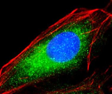
ARG58289 anti-RPL4 antibody ICC/IF image
Immunofluorescence: 4% paraformaldehyde-fixed, 0.1% Triton X-100 permeabilized U-2 OS cells stained with ARG58289 anti-RPL4 antibody (green) at 1:25 dilution. Cytoplasmic actin is detected with Dylight® 554 Phalloidin at 1:100 dilution (red). DAPI (blue) for nuclear staining.
-
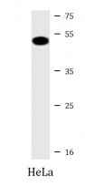
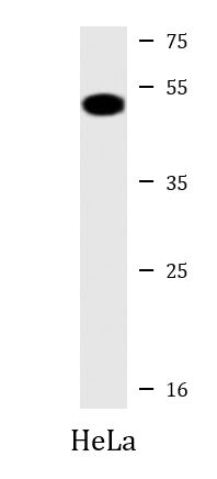
ARG58289 anti-RPL4 antibody WB image
Western blot: 20 µg of HeLa cell lysate stained with ARG58289 anti-RPL4 antibody at 1:2000 dilution.
-
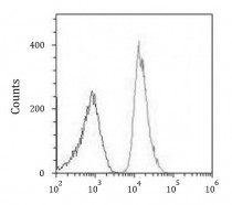
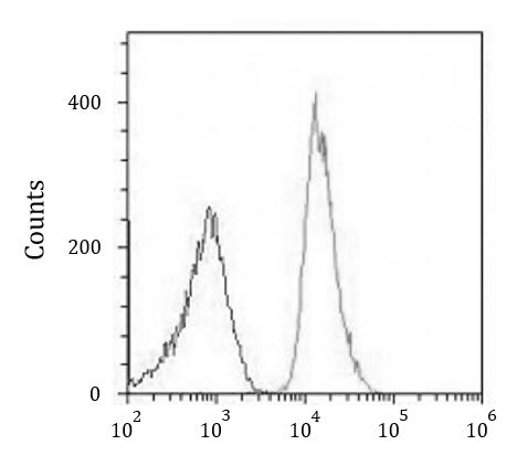
ARG58289 anti-RPL4 antibody FACS image
Flow Cytometry: U-2OS cells were fixed with 2% paraformaldehyde (10 min) and then permeabilized with 90% methanol for 10 min. The cells were then incubated in 2% BSA to block non-specific protein-protein interactions and stained with ARG58289 anti-RPL4 antibody (right histogram) at 1:25 dilution for 60 min at 37°C, followed by incubation with DyLight® 488 labelled secondary antibody. Isotype control antibody (left histogram) was Rabbit IgG (1 µg/10^6 cells) used under the same conditions. Acquisition of >10000 events was performed.