ARG43356
anti-SARS-CoV-2 NSP14 ExoN antibody
anti-SARS-CoV-2 NSP14 ExoN antibody for ELISA,IHC-Formalin-fixed paraffin-embedded sections and Virus
Overview
| Product Description | Rabbit Polyclonal antibody recognizes SARS-CoV-2 NSP14 ExoN |
|---|---|
| Tested Reactivity | Virus |
| Tested Application | ELISA, IHC-P |
| Host | Rabbit |
| Clonality | Polyclonal |
| Isotype | IgG |
| Target Name | SARS-CoV-2 NSP14 ExoN |
| Antigen Species | Virus |
| Immunogen | A 14-amino acid synthetic peptide between aa. 120-170 of SARS-CoV-2 NSP14 ExoN. |
| Conjugation | Un-conjugated |
Application Instructions
| Application Suggestion |
|
||||||
|---|---|---|---|---|---|---|---|
| Application Note | IHC-P: Antigen Retrieval: Heat mediation was performed in Citrate buffer (pH 6.0). * The dilutions indicate recommended starting dilutions and the optimal dilutions or concentrations should be determined by the scientist. |
Properties
| Form | Liquid |
|---|---|
| Purification | Affinity purification with immunogen. |
| Buffer | PBS and 0.02% Sodium azide. |
| Preservative | 0.02% Sodium azide |
| Concentration | 1 mg/ml |
| Storage Instruction | For continuous use, store undiluted antibody at 2-8°C for up to a week. For long-term storage, aliquot and store at -20°C or below. Storage in frost free freezers is not recommended. Avoid repeated freeze/thaw cycles. Suggest spin the vial prior to opening. The antibody solution should be gently mixed before use. |
| Note | For laboratory research only, not for drug, diagnostic or other use. |
Images (1) Click the Picture to Zoom In
-
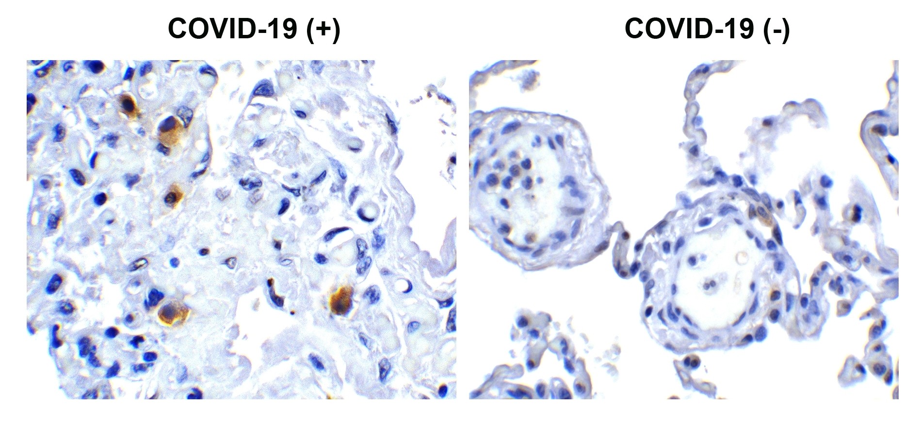
ARG43356 anti-SARS-CoV-2 NSP14 ExoN antibody IHC-P image
Immunohistochemistry: Formaldehyde-fixed and paraffin-embedded COVID-19 patient lung tissue. Tissue was blocked with 10% serum for 1 hour at RT. Antigen Retrieval: Heat mediation was performed in Citrate buffer (pH 6.0). Samples were stained with ARG43356 anti-SARS-CoV-2 NSP14 ExoN antibody at 0.5 µg/ml dilution, overnight at 4°C. Counter stained with Hematoxylin. Strong signal of SARS-COV-2 NSP14 protein was observed in macrophage of COVID-19 patient lung, but not in non-COVID-19 patient lung.