ARG54885
anti-SARS-CoV Spike protein antibody
anti-SARS-CoV Spike protein antibody for Western blot and Virus
Microbiology and Infectious Disease antibody
Overview
| Product Description | Rabbit Polyclonal antibody recognizes SARS-CoV Spike protein. |
|---|---|
| Tested Reactivity | Virus |
| Tested Application | WB |
| Host | Rabbit |
| Clonality | Polyclonal |
| Isotype | IgG |
| Target Name | SARS-CoV Spike protein |
| Immunogen | KLH-conjugated synthetic peptide corresponding to aa. 532-562 of the middle of SARS-CoV Spike protein. |
| Conjugation | Un-conjugated |
Application Instructions
| Application Suggestion |
|
||||
|---|---|---|---|---|---|
| Application Note | * The dilutions indicate recommended starting dilutions and the optimal dilutions or concentrations should be determined by the scientist. |
Properties
| Form | Liquid |
|---|---|
| Purification | Purification with Protein G. |
| Buffer | PBS and 0.09% (W/V) Sodium azide |
| Preservative | 0.09% (W/V) Sodium azide |
| Storage Instruction | For continuous use, store undiluted antibody at 2-8°C for up to a week. For long-term storage, aliquot and store at -20°C or below. Storage in frost free freezers is not recommended. Avoid repeated freeze/thaw cycles. Suggest spin the vial prior to opening. The antibody solution should be gently mixed before use. |
| Note | For laboratory research only, not for drug, diagnostic or other use. |
Bioinformation
| Function | S1 attaches the virion to the cell membrane by interacting with human ACE2 and CLEC4M/DC-SIGNR, initiating the infection. Binding to the receptor and internalization of the virus into the endosomes of the host cell probably induces conformational changes in the S glycoprotein. Proteolysis by cathepsin CTSL may unmask the fusion peptide of S2 and activate membranes fusion within endosomes. S2 is a class I viral fusion protein. Under the current model, the protein has at least three conformational states: pre-fusion native state, pre-hairpin intermediate state, and post-fusion hairpin state. During viral and target cell membrane fusion, the coiled coil regions (heptad repeats) assume a trimer-of-hairpins structure, positioning the fusion peptide in close proximity to the C-terminal region of the ectodomain. The formation of this structure appears to drive apposition and subsequent fusion of viral and target cell membranes. [UniProt] |
|---|---|
| Cellular Localization | Virion membrane; Single-pass type I membrane protein. Host endoplasmic reticulum-Golgi intermediate compartment membrane; Single-pass type I membrane protein. Host cell membrane; Single-pass type I membrane protein. Note=Accumulates in the endoplasmic reticulum-Golgi intermediate compartment, where it participates in virus particle assembly (By similarity). Some S oligomers are transported to the plasma membrane, where they may mediate cell- cell fusion. |
| Highlight | Related products: SARS-CoV antibodies; SARS-CoV ELISA Kits; SARS-CoV recombinant proteins; Anti-Rabbit IgG secondary antibodies; Related news: HMGB1, a biomarker and therapeutic target in COVID-19 ACE2, receptor of 2019-nCoV Exploring Antiviral Immune Response |
| Research Area | Microbiology and Infectious Disease antibody |
Images (1) Click the Picture to Zoom In
-
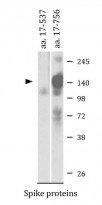
ARG54885 anti-SARS-CoV Spike protein antibody WB image
Western blot: Recombinant Spike proteins (aa. 17-537 or aa. 17-756) stained with ARG54885 anti-SARS-CoV Spike protein antibody.

Specific References
Assessing the application of a pseudovirus system for emerging SARS-CoV-2 and re-emerging avian influenza virus H5 subtypes in vaccine development.
WB / Virus


