ARG63204
anti-Septin 3 antibody
anti-Septin 3 antibody for IHC-Formalin-fixed paraffin-embedded sections,Western blot and Human
Cell Biology and Cellular Response antibody
Overview
| Product Description | Goat Polyclonal antibody recognizes Septin 3 |
|---|---|
| Tested Reactivity | Hu |
| Predict Reactivity | Ms, Rat, Cow, Dog, Pig |
| Tested Application | IHC-P, WB |
| Specificity | This antibody is expected to recognize both reported isoforms according to NP_663786.1 and NP_061979.3. |
| Host | Goat |
| Clonality | Polyclonal |
| Isotype | IgG |
| Target Name | Septin 3 |
| Antigen Species | Human |
| Immunogen | SELVPEPRPKPA-C |
| Conjugation | Un-conjugated |
| Alternate Names | SEP3; Neuronal-specific septin-3; bK250D10.3 |
Application Instructions
| Application Suggestion |
|
||||||
|---|---|---|---|---|---|---|---|
| Application Note | WB: Recommend incubate at RT for 1h. IHC-P: Antigen Retrieval: Steam tissue section in Citrate buffer (pH 6.0). * The dilutions indicate recommended starting dilutions and the optimal dilutions or concentrations should be determined by the scientist. |
Properties
| Form | Liquid |
|---|---|
| Purification | Purified from goat serum by antigen affinity chromatography. |
| Buffer | Tris saline (pH 7.3), 0.02% Sodium azide and 0.5% BSA. |
| Preservative | 0.02% Sodium azide |
| Stabilizer | 0.5% BSA |
| Concentration | 0.5 mg/ml |
| Storage Instruction | For continuous use, store undiluted antibody at 2-8°C for up to a week. For long-term storage, aliquot and store at -20°C or below. Storage in frost free freezers is not recommended. Avoid repeated freeze/thaw cycles. Suggest spin the vial prior to opening. The antibody solution should be gently mixed before use. |
| Note | For laboratory research only, not for drug, diagnostic or other use. |
Bioinformation
| Database Links | |
|---|---|
| Background | This gene belongs to the septin family of GTPases. Members of this family are required for cytokinesis. Expression is upregulated by retinoic acid in a human teratocarcinoma cell line. The specific function of this gene has not been determined. Alternative splicing of this gene results in two transcript variants encoding different isoforms. [provided by RefSeq, Jul 2008] |
| Research Area | Cell Biology and Cellular Response antibody |
| Calculated MW | 41 kDa |
| PTM | Phosphorylated by PKG on serine residues. Phosphorylated by PKG on Ser-91 (By similarity). |
Images (4) Click the Picture to Zoom In
-
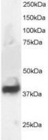
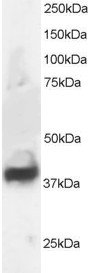
ARG63204 anti-Septin 3 antibody WB image
Western Blot: Human Brain lysate (RIPA buffer, 30µg total protein per lane) stained with ARG63204 anti-Septin 3 antibody at 1 µg/ml dilution.
-
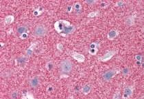
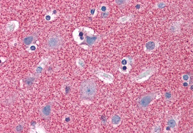
ARG63204 anti-Septin 3 antibody IHC-P image
Immunohistochemistry: Paraffin-embedded Human cortex tissue. Antigen Retrieval: Steam tissue section in Citrate buffer (pH 6.0). The tissue section was stained with ARG63204 anti-Septin 3 antibody at 3.75 µg/ml dilution followed by AP-staining.
-
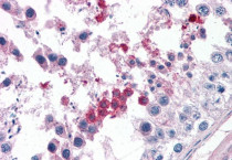
ARG63204 anti-Septin 3 antibody IHC-P image
Immunohistochemistry: Paraffin-embedded Human testis tissue. Antigen Retrieval: Steam tissue section in Citrate buffer (pH 6.0). The tissue section was stained with ARG63204 anti-Septin 3 antibody at 3.75 µg/ml dilution followed by AP-staining.
-
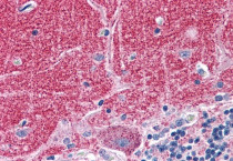
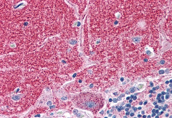
ARG63204 anti-Septin 3 antibody IHC-P image
Immunohistochemistry: Paraffin-embedded Human cerebellum tissue. Antigen Retrieval: Steam tissue section in Citrate buffer (pH 6.0). The tissue section was stained with ARG63204 anti-Septin 3 antibody at 3.75 µg/ml dilution followed by AP-staining.