ARG43065
anti-TXNIP antibody
anti-TXNIP antibody for Flow cytometry,IHC-Formalin-fixed paraffin-embedded sections,Western blot and Human,Mouse,Rat
Overview
| Product Description | Rabbit Polyclonal antibody recognizes TXNIP |
|---|---|
| Tested Reactivity | Hu, Ms, Rat |
| Tested Application | FACS, IHC-P, WB |
| Host | Rabbit |
| Clonality | Polyclonal |
| Isotype | IgG |
| Target Name | TXNIP |
| Antigen Species | Human |
| Immunogen | Recombinant protein corresponding to K5-Q391 of Human TXNIP. |
| Conjugation | Un-conjugated |
| Alternate Names | Thioredoxin-binding protein 2; Vitamin D3 up-regulated protein 1; Thioredoxin-interacting protein; HHCPA78; VDUP1; THIF; EST01027 |
Application Instructions
| Application Suggestion |
|
||||||||
|---|---|---|---|---|---|---|---|---|---|
| Application Note | IHC-P: Antigen Retrieval: Heat mediation was performed in EDTA buffer (pH 8.0). * The dilutions indicate recommended starting dilutions and the optimal dilutions or concentrations should be determined by the scientist. |
Properties
| Form | Liquid |
|---|---|
| Purification | Affinity purification with immunogen. |
| Buffer | 0.2% Na2HPO4, 0.9% NaCl, 0.05% Sodium azide and 4% Trehalose. |
| Preservative | 0.05% Sodium azide |
| Stabilizer | 4% Trehalose |
| Concentration | 0.5 mg/ml |
| Storage Instruction | For continuous use, store undiluted antibody at 2-8°C for up to a week. For long-term storage, aliquot and store at -20°C or below. Storage in frost free freezers is not recommended. Avoid repeated freeze/thaw cycles. Suggest spin the vial prior to opening. The antibody solution should be gently mixed before use. |
| Note | For laboratory research only, not for drug, diagnostic or other use. |
Bioinformation
| Database Links | |
|---|---|
| Gene Symbol | TXNIP |
| Gene Full Name | thioredoxin interacting protein |
| Background | This gene encodes a thioredoxin-binding protein that is a member of the alpha arrestin protein family. Thioredoxin is a thiol-oxidoreductase that is a major regulator of cellular redox signaling which protects cells from oxidative stress. This protein inhibits the antioxidative function of thioredoxin resulting in the accumulation of reactive oxygen species and cellular stress. This protein also functions as a regulator of cellular metabolism and of endoplasmic reticulum (ER) stress. This protein may also function as a tumor suppressor. Alternate splicing results in multiple transcript variants. [provided by RefSeq, Sep 2015] |
| Function | May act as an oxidative stress mediator by inhibiting thioredoxin activity or by limiting its bioavailability. Interacts with COPS5 and restores COPS5-induced suppression of CDKN1B stability, blocking the COPS5-mediated translocation of CDKN1B from the nucleus to the cytoplasm. Functions as a transcriptional repressor, possibly by acting as a bridge molecule between transcription factors and corepressor complexes, and over-expression will induce G0/G1 cell cycle arrest. Required for the maturation of natural killer cells. Acts as a suppressor of tumor cell growth. Inhibits the proteasomal degradation of DDIT4, and thereby contributes to the inhibition of the mammalian target of rapamycin complex 1 (mTORC1). [UniProt] |
| Cellular Localization | Cytoplasm. [UniProt] |
| Calculated MW | 44 kDa |
| PTM | Ubiquitinated; undergoes polyubiquitination catalyzed by ITCH resulting in proteasomal degradation. [UniProt] |
Images (3) Click the Picture to Zoom In
-
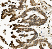
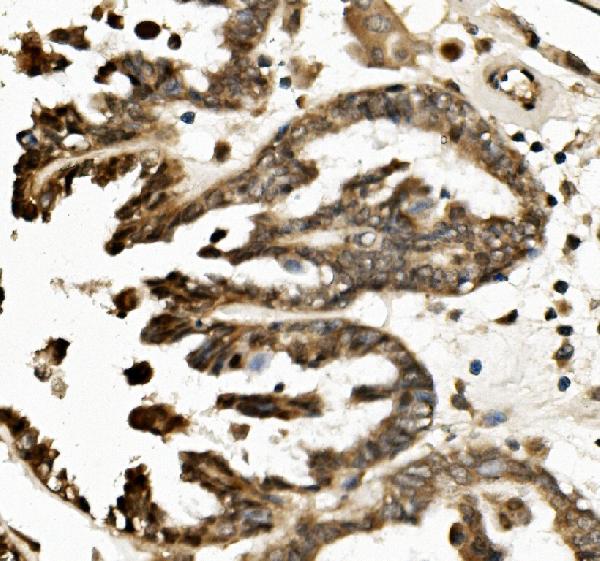
ARG43065 anti-TXNIP antibody IHC-P image
Immunohistochemistry: Paraffin-embedded Human ovarian cancer tissue. Antigen Retrieval: Heat mediation was performed in EDTA buffer (pH 8.0). The tissue section was blocked with 10% goat serum. The tissue section was then stained with ARG43065 anti-TXNIP antibody at 1 µg/ml dilution, overnight at 4°C.
-
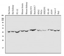
ARG43065 anti-TXNIP antibody WB image
Western blot: 50 µg of sample under reducing conditions. Rat brain, Rat kidney, PC-12, Mouse brain, Mouse kidney, NIH/3T3, Raw264.7, K562, HeLa, HEK293, HL-60, A549, Caco-2 and Raji whole cell lysates stained with ARG43065 anti-TXNIP antibody at 0.5 µg/ml dilution, overnight at 4°C.
-
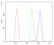
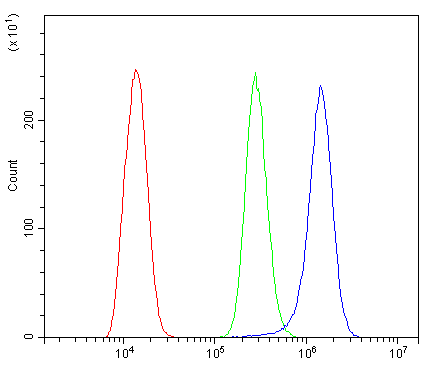
ARG43065 anti-TXNIP antibody FACS image
Flow Cytometry: THP-1 cells were blocked with 10% normal goat serum and then stained with ARG43065 anti-TXNIP antibody (blue) at 1 µg/10^6 cells for 30 min at 20°C, followed by incubation with DyLight®488 labelled secondary antibody. Isotype control antibody (green) was rabbit IgG (1 µg/10^6 cells) used under the same conditions. Unlabelled sample (red) was also used as a control.