ARG52470
anti-Visinin like 1 antibody [2D11]
anti-Visinin like 1 antibody [2D11] for ICC/IF,IHC-Frozen sections,Western blot and Human,Mouse,Rat,Bovine
Cancer antibody; Signaling Transduction antibody
Overview
| Product Description | Mouse Monoclonal antibody [2D11] recognizes Visinin like 1 |
|---|---|
| Tested Reactivity | Hu, Ms, Rat, Bov |
| Tested Application | ICC/IF, IHC-Fr, WB |
| Host | Mouse |
| Clonality | Monoclonal |
| Clone | 2D11 |
| Isotype | IgG1 |
| Target Name | Visinin like 1 |
| Antigen Species | Human |
| Immunogen | Recombinant human VSNL1 purified from E. coli |
| Conjugation | Un-conjugated |
| Alternate Names | HPCAL3; HUVISL1; Visinin-like protein 1; HLP3; Hippocalcin-like protein 3; VILIP; VLP-1; VILIP-1 |
Application Instructions
| Application Suggestion |
|
||||||||
|---|---|---|---|---|---|---|---|---|---|
| Application Note | Specific for the ~22k protein * The dilutions indicate recommended starting dilutions and the optimal dilutions or concentrations should be determined by the scientist. |
Properties
| Form | Liquid |
|---|---|
| Purification | Affinity Purified |
| Buffer | PBS and 10 mM Sodium azide |
| Preservative | 10 mM Sodium azide |
| Storage Instruction | For continuous use, store undiluted antibody at 2-8°C for up to a week. For long-term storage, aliquot and store at -20°C or below. Storage in frost free freezers is not recommended. Avoid repeated freeze/thaw cycles. Suggest spin the vial prior to opening. The antibody solution should be gently mixed before use. |
| Note | For laboratory research only, not for drug, diagnostic or other use. |
Bioinformation
| Database Links | |
|---|---|
| Gene Symbol | VSNL1 |
| Gene Full Name | visinin-like 1 |
| Background | Visinin-like protein 1 (VSNL1), also known as VILIP1, is a calcium sensor protein expressed exclusively in neurons. Highest levels of VSNL1 expression are found in cerebellar Purkinje cells. VSNL1 has been implicated in the modulation of cell signaling cascades via regulation of adenyl cyclase activity (Braunewell et al., 1997). Additionally, VSNL1 has been associated with amyloid plaques and neurofibrillar tangles in Alzheimer’s disease (Schnurra et al., 2001). |
| Research Area | Cancer antibody; Signaling Transduction antibody |
| Calculated MW | 22 kDa |
Images (4) Click the Picture to Zoom In
-
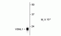
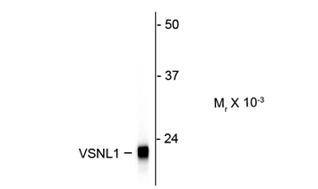
ARG52470 anti-Visinin like 1 antibody [2D11] WB image
Western blot: Rat cerebellum lysate showing specific immunolabeling of the ~ 22k VSNL1 protein stained with ARG52470 anti-Visinin like 1 antibody [2D11].
-

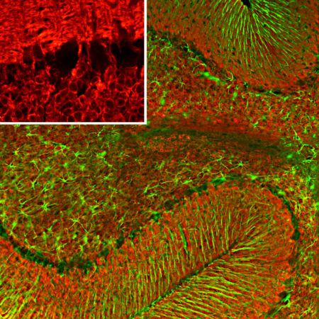
ARG52470 anti-Visinin like 1 antibody [2D11] IHC-Fr image
Immunohistochemistry: Frozen section of Rat cerebellum tissue stained with ARG52470 anti-Visinin like 1 antibody [2D11] (red) at 1:500 dilution, and costained with anti-GFAP antibody (green) at 1:5000 dilution. DAPI (blue) for nuclear staining. Following transcardial perfusion of Rat with 4% paraformaldehyde, brain was post fixed for 24 hours, cut to 45 µM, and free-floating sections were stained with the above antibodies.
Clone 2D11 reveals protein expressed in granule cell membranes and in synapses in the white matter and molecular layers of the cerebellum. The GFAP antibody stains the processes of Bergmann glia and astroglia.
-
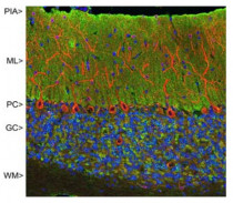
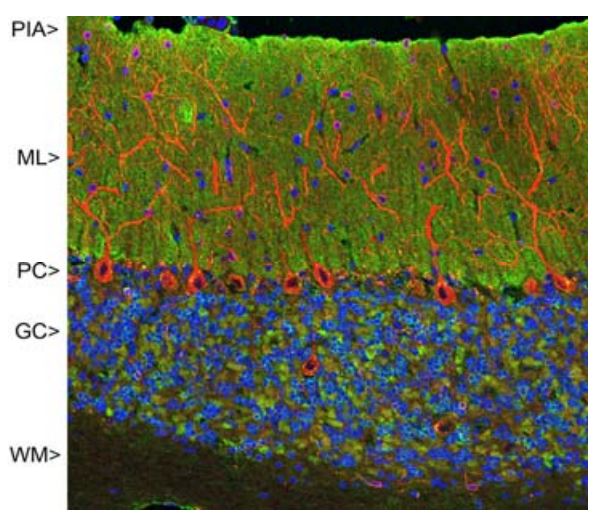
ARG52470 anti-Visinin like 1 antibody [2D11] IHC image
Immunohistochemistry: Rat cerebellum stained with ARG52470 anti-Visinin like 1 antibody [2D11] showing strong synaptic staining of VSNL1 (green) in the molecular layer (ML) and MAP2 stained with ARG52328 anti-MAP2 antibody in red.
-

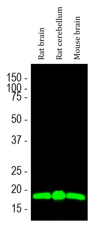
ARG52470 anti-Visinin like 1 antibody [2D11] WB image
Western blot: Rat brain, Rat cerebellum and Mouse brain lysates stained with ARG52470 anti-Visinin like 1 antibody [2D11] (green) at 1:1000 dilution.