ARG42701
anti-DIO1 antibody
anti-DIO1 antibody for Flow cytometry,ICC/IF,Western blot and Human,Mouse,Rat
Overview
| Product Description | Rabbit Polyclonal antibody recognizes DIO1 |
|---|---|
| Tested Reactivity | Hu, Ms, Rat |
| Tested Application | FACS, ICC/IF, WB |
| Host | Rabbit |
| Clonality | Polyclonal |
| Isotype | IgG |
| Target Name | DIO1 |
| Antigen Species | Human |
| Immunogen | Recombinant protein corresponding to K137-S249 of Human DIO1. |
| Conjugation | Un-conjugated |
| Alternate Names | EC 1.97.1.10; Type I iodothyronine deiodinase; TXDI1; DIOI; 5DI; Type-I 5'-deiodinase; Type 1 DI |
Application Instructions
| Application Suggestion |
|
||||||||
|---|---|---|---|---|---|---|---|---|---|
| Application Note | * The dilutions indicate recommended starting dilutions and the optimal dilutions or concentrations should be determined by the scientist. | ||||||||
| Observed Size | ~ 29 kDa |
Properties
| Form | Liquid |
|---|---|
| Purification | Affinity purification with immunogen. |
| Buffer | 0.2% Na2HPO4, 0.9% NaCl, 0.01% Sodium azide and 4% Trehalose. |
| Preservative | 0.01% Sodium azide |
| Stabilizer | 4% Trehalose |
| Concentration | 0.5 mg/ml |
| Storage Instruction | For continuous use, store undiluted antibody at 2-8°C for up to a week. For long-term storage, aliquot and store at -20°C or below. Storage in frost free freezers is not recommended. Avoid repeated freeze/thaw cycles. Suggest spin the vial prior to opening. The antibody solution should be gently mixed before use. |
| Note | For laboratory research only, not for drug, diagnostic or other use. |
Bioinformation
| Database Links | |
|---|---|
| Gene Symbol | DIO1 |
| Gene Full Name | deiodinase, iodothyronine, type I |
| Background | The protein encoded by this gene belongs to the iodothyronine deiodinase family. It catalyzes the activation, as well as the inactivation of thyroid hormone by outer and inner ring deiodination, respectively. The activation reaction involves the conversion of the prohormone thyroxine (3,5,3',5'-tetraiodothyronine, T4), secreted by the thyroid gland, to the bioactive thyroid hormone (3,5,3'-triiodothyronine, T3) by 5'-deiodination. This protein provides most of the circulating T3, which is essential for growth, differentiation and basal metabolism in vertebrates. This protein is a selenoprotein, containing the rare amino acid selenocysteine (Sec) at its active site. Sec is encoded by the UGA codon, which normally signals translation termination. The 3' UTRs of selenoprotein mRNAs contain a conserved stem-loop structure, designated the Sec insertion sequence (SECIS) element, that is necessary for the recognition of UGA as a Sec codon, rather than as a stop signal. Alternatively spliced transcript variants have been found for this gene. [provided by RefSeq, Jun 2018] |
| Function | Responsible for the deiodination of T4 (3,5,3',5'-tetraiodothyronine) into T3 (3,5,3'-triiodothyronine) and of T3 into T2 (3,3'-diiodothyronine). Plays a role in providing a source of plasma T3 by deiodination of T4 in peripheral tissues such as liver and kidney. [UniProt] |
| Cellular Localization | Endoplasmic reticulum membrane; Single-pass membrane protein. [UniProt] |
| Calculated MW | 29 kDa |
Images (4) Click the Picture to Zoom In
-
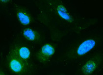
ARG42701 anti-DIO1 antibody ICC/IF image
Immunofluorescence: A549 cells were blocked with 10% goat serum and then stained with ARG42701 anti-DIO1 antibody (green) at 2 µg/ml dilution, overnight at 4°C. DAPI (blue) for nuclear staining.
-
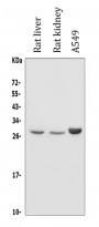
ARG42701 anti-DIO1 antibody WB image
Western blot: 50 µg of samples under reducing condition. Rat liver, Rat kidney and A549 whole cell lysates stained with ARG42701 anti-DIO1 antibody at 0.5 µg/ml dilution, overnight at 4°C.
-

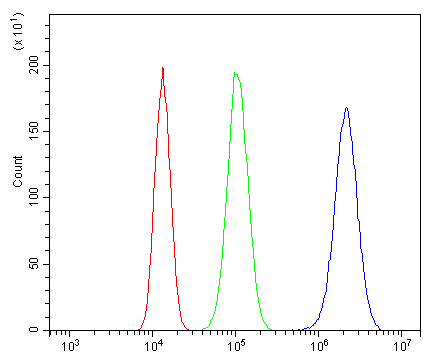
ARG42701 anti-DIO1 antibody FACS image
Flow Cytometry: 293T cells were blocked with 10% normal goat serum and then stained with ARG42701 anti-DIO1 antibody (blue) at 1 µg/10^6 cells for 30 min at 20°C, followed by incubation with DyLight®488 labelled secondary antibody. Isotype control antibody (green) was rabbit IgG (1 µg/10^6 cells) used under the same conditions. Unlabelled sample (red) was also used as a control.
-

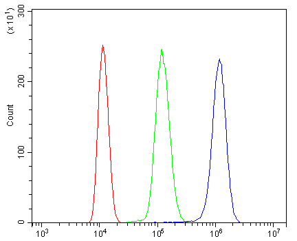
ARG42701 anti-DIO1 antibody FACS image
Flow Cytometry: ANA-1 cells were blocked with 10% normal goat serum and then stained with ARG42701 anti-DIO1 antibody (blue) at 1 µg/10^6 cells for 30 min at 20°C, followed by incubation with DyLight®488 labelled secondary antibody. Isotype control antibody (green) was rabbit IgG (1 µg/10^6 cells) used under the same conditions. Unlabelled sample (red) was also used as a control.