ARG41529
anti-DMD / Dystrophin antibody
anti-DMD / Dystrophin antibody for Western blot and Human
Overview
| Product Description | Rabbit Polyclonal antibody recognizes DMD / Dystrophin |
|---|---|
| Tested Reactivity | Hu |
| Predict Reactivity | Ms, Rat |
| Tested Application | WB |
| Host | Rabbit |
| Clonality | Polyclonal |
| Isotype | IgG |
| Target Name | DMD / Dystrophin |
| Antigen Species | Human |
| Immunogen | Synthetic peptide of Human DMD / Dystrophin. |
| Conjugation | Un-conjugated |
| Alternate Names | BMD; CMD3B; DXS270; DXS272; MRX85; Dystrophin; DXS164; DXS239; DXS206; DXS142; DXS230; DXS269; DXS268 |
Application Instructions
| Application Suggestion |
|
||||
|---|---|---|---|---|---|
| Application Note | * The dilutions indicate recommended starting dilutions and the optimal dilutions or concentrations should be determined by the scientist. | ||||
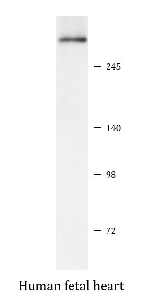
| Positive Control | Human fetal heart |
Properties
| Form | Liquid |
|---|---|
| Purification | Affinity purified. |
| Buffer | PBS (pH 7.4), 150 mM NaCl, 0.02% Sodium azide and 50% Glycerol. |
| Preservative | 0.02% Sodium azide |
| Stabilizer | 50% Glycerol |
| Storage Instruction | For continuous use, store undiluted antibody at 2-8°C for up to a week. For long-term storage, aliquot and store at -20°C. Storage in frost free freezers is not recommended. Avoid repeated freeze/thaw cycles. Suggest spin the vial prior to opening. The antibody solution should be gently mixed before use. |
| Note | For laboratory research only, not for drug, diagnostic or other use. |
Bioinformation
| Database Links | |
|---|---|
| Gene Symbol | DMD |
| Gene Full Name | dystrophin |
| Background | The dystrophin gene is the largest gene found in nature, measuring 2.4 Mb. The gene was identified through a positional cloning approach, targeted at the isolation of the gene responsible for Duchenne (DMD) and Becker (BMD) Muscular Dystrophies. DMD is a recessive, fatal, X-linked disorder occurring at a frequency of about 1 in 3,500 new-born males. BMD is a milder allelic form. In general, DMD patients carry mutations which cause premature translation termination (nonsense or frame shift mutations), while in BMD patients dystrophin is reduced either in molecular weight (derived from in-frame deletions) or in expression level. The dystrophin gene is highly complex, containing at least eight independent, tissue-specific promoters and two polyA-addition sites. Furthermore, dystrophin RNA is differentially spliced, producing a range of different transcripts, encoding a large set of protein isoforms. Dystrophin (as encoded by the Dp427 transcripts) is a large, rod-like cytoskeletal protein which is found at the inner surface of muscle fibers. Dystrophin is part of the dystrophin-glycoprotein complex (DGC), which bridges the inner cytoskeleton (F-actin) and the extra-cellular matrix. [provided by RefSeq, Jul 2008] |
| Function | Anchors the extracellular matrix to the cytoskeleton via F-actin. Ligand for dystroglycan. Component of the dystrophin-associated glycoprotein complex which accumulates at the neuromuscular junction (NMJ) and at a variety of synapses in the peripheral and central nervous systems and has a structural function in stabilizing the sarcolemma. Also implicated in signaling events and synaptic transmission. [UniProt] |
| Cellular Localization | Cell membrane, sarcolemma; Peripheral membrane protein; Cytoplasmic side. Cytoplasm, cytoskeleton. Cell junction, synapse, postsynaptic cell membrane. Note=In muscle cells, sarcolemma localization requires the presence of ANK2, while localization to costameres requires the presence of ANK3. Localizes to neuromuscular junctions (NMJs). In adult muscle, NMJ localization depends upon ANK2 presence, but not in newborn animals. [UniProt] |
| Calculated MW | 427 kDa |
Images (1) Click the Picture to Zoom In
-
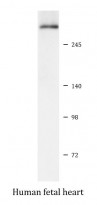
ARG41529 anti-DMD / Dystrophin antibody WB image
Western blot: Human fetal heart lysate stained with ARG41529 anti-DMD / Dystrophin antibody.