ARG41755
anti-GLUT1 antibody [GLUT1/2475]
anti-GLUT1 antibody [GLUT1/2475] for Flow cytometry,ICC/IF,IHC-Formalin-fixed paraffin-embedded sections,Western blot and Human
Overview
| Product Description | Mouse Monoclonal antibody [GLUT1/2475] recognizes GLUT1 |
|---|---|
| Tested Reactivity | Hu |
| Tested Application | FACS, ICC/IF, IHC-P, WB |
| Host | Mouse |
| Clonality | Monoclonal |
| Clone | GLUT1/2475 |
| Isotype | IgG2b, kappa |
| Target Name | GLUT1 |
| Antigen Species | Human |
| Immunogen | Synthetic peptide corresponding to aa. 203-305 of Human GLUT1. |
| Conjugation | Un-conjugated |
| Alternate Names | DYT17; HepG2 glucose transporter; CSE; GLUT-1; GLUT; GLUT1DS; DYT18; HTLVR; PED; Glucose transporter type 1, erythrocyte/brain; DYT9; EIG12; GLUT1; Solute carrier family 2, facilitated glucose transporter member 1 |
Application Instructions
| Application Suggestion |
|
||||||||||
|---|---|---|---|---|---|---|---|---|---|---|---|
| Application Note | IHC-P: Antigen Retrieval: Boil tissue section in 10 mM Tris with 1 mM EDTA (pH 9.0) for 10-20 min, followed by cooling at RT for 20 min. * The dilutions indicate recommended starting dilutions and the optimal dilutions or concentrations should be determined by the scientist. |
Properties
| Form | Liquid |
|---|---|
| Purification | Purification with Protein G. |
| Buffer | PBS, 0.05% Sodium azide and 0.1 mg/ml BSA. |
| Preservative | 0.05% Sodium azide |
| Stabilizer | 0.1 mg/ml BSA |
| Concentration | 0.2 mg/ml |
| Storage Instruction | For continuous use, store undiluted antibody at 2-8°C for up to a week. For long-term storage, aliquot and store at -20°C or below. Storage in frost free freezers is not recommended. Avoid repeated freeze/thaw cycles. Suggest spin the vial prior to opening. The antibody solution should be gently mixed before use. |
| Note | For laboratory research only, not for drug, diagnostic or other use. |
Bioinformation
| Database Links |
Swiss-port # P11166 Human Solute carrier family 2, facilitated glucose transporter member 1 |
|---|---|
| Gene Symbol | SLC2A1 |
| Gene Full Name | solute carrier family 2 (facilitated glucose transporter), member 1 |
| Background | This gene encodes a major glucose transporter in the mammalian blood-brain barrier. The encoded protein is found primarily in the cell membrane and on the cell surface, where it can also function as a receptor for human T-cell leukemia virus (HTLV) I and II. Mutations in this gene have been found in a family with paroxysmal exertion-induced dyskinesia. [provided by RefSeq, Apr 2013] |
| Function | Facilitative glucose transporter. This isoform may be responsible for constitutive or basal glucose uptake. Has a very broad substrate specificity; can transport a wide range of aldoses including both pentoses and hexoses. [UniProt] |
| Cellular Localization | Cell membrane; Multi-pass membrane protein. Melanosome. Note=Localizes primarily at the cell surface. Identified by mass spectrometry in melanosome fractions from stage I to stage IV. [UniProt] |
| Calculated MW | 54 kDa |
Images (5) Click the Picture to Zoom In
-
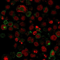
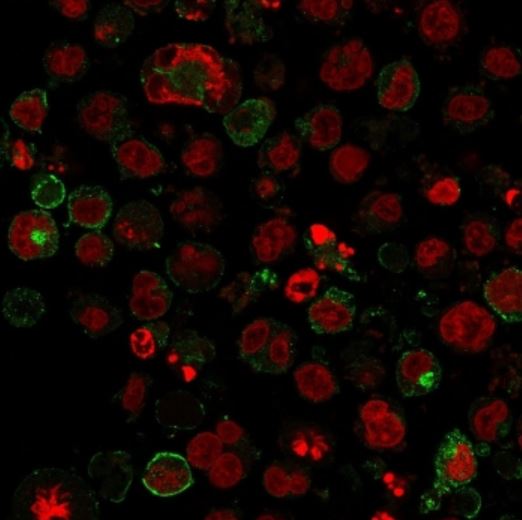
ARG41755 anti-GLUT1 antibody [GLUT1/2475] ICC/IF image
Immunofluorescence: K562 cells stained with ARG41755 anti-GLUT1 antibody [GLUT1/2475] (green). Reddot (red) for nuclear staining.
-
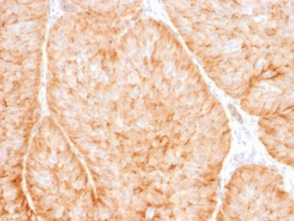
ARG41755 anti-GLUT1 antibody [GLUT1/2475] IHC-P image
Immunohistochemistry: Paraffin-embedded Human bladder carcinoma tissue. Antigen Retrieval: Boil tissue section in 10 mM Tris with 1 mM EDTA (pH 9.0) for 10-20 min, followed by cooling at RT for 20 min. The tissue section was stained with ARG41755 anti-GLUT1 antibody [GLUT1/2475].
-
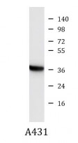
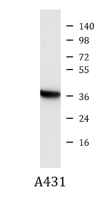
ARG41755 anti-GLUT1 antibody [GLUT1/2475] WB image
Western blot: A431 cell lysate stained with ARG41755 anti-GLUT1 antibody [GLUT1/2475].
-
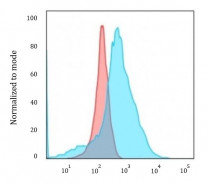
ARG41755 anti-GLUT1 antibody [GLUT1/2475] FACS image
Flow Cytometry: K562 cells stained with ARG41755 anti-GLUT1 antibody [GLUT1/2475] (blue) or isotype control antibody (red), followed by incubation with FITC labelled secondary antibody.
-

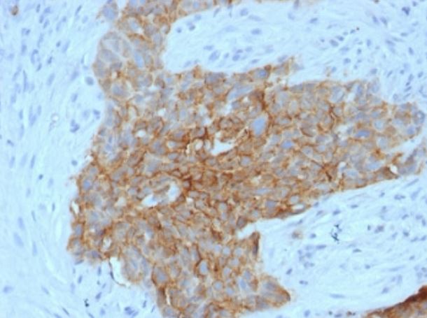
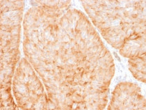
ARG41755 anti-GLUT1 antibody [GLUT1/2475] IHC-P image
Immunohistochemistry: Paraffin-embedded human tongue tissue. Antigen Retrieval: Boil tissue section in 10 mM Tris with 1 mM EDTA (pH 9.0) for 10-20 min, followed by cooling at RT for 20 min. The tissue section was stained with ARG41755 anti-GLUT1 antibody [GLUT1/2475].