ARG43932
anti-PKC alpha antibody
anti-PKC alpha antibody for ELISA,Flow cytometry,Western blot and Human,Mouse,Rat
Overview
| Product Description | Rabbit Polyclonal antibody recognizes PKC alpha |
|---|---|
| Tested Reactivity | Hu, Ms, Rat |
| Tested Application | ELISA, FACS, WB |
| Host | Rabbit |
| Clonality | Polyclonal |
| Isotype | IgG |
| Target Name | PKC alpha |
| Antigen Species | Human |
| Immunogen | Human PKC alpha recombinant protein |
| Expression System | E.coli |
| Conjugation | Un-conjugated |
| Protein Full Name | Protein kinase C alpha type |
| Alternate Names | PRKCA; Protein Kinase C Alpha; PKCA; Protein Kinase C Alpha Type; EC 2.7.11.13; PKC-Alpha; PKCalpha; PRKACA; PKC-A; Protein Kinase C, Alpha; Aging-Associated Gene 6; EC 2.7.11; PKCI+/-; PKCα; AAG6 |
Application Instructions
| Application Suggestion |
|
||||||||
|---|---|---|---|---|---|---|---|---|---|
| Application Note | * The dilutions indicate recommended starting dilutions and the optimal dilutions or concentrations should be determined by the scientist. |
Properties
| Form | Liquid |
|---|---|
| Purification | Affinity purified with Immunogen. |
| Buffer | 0.9% NaCl, 0.2% Na2HPO4 and 4% Trehalose. |
| Stabilizer | 4% Trehalose |
| Concentration | 0.5 mg/ml |
| Storage Instruction | For continuous use, store undiluted antibody at 2-8°C for up to a week. For long-term storage, aliquot and store at -20°C or below. Storage in frost free freezers is not recommended. Avoid repeated freeze/thaw cycles. Suggest spin the vial prior to opening. The antibody solution should be gently mixed before use. |
| Note | For laboratory research only, not for drug, diagnostic or other use. |
Bioinformation
| Database Links | |
|---|---|
| Gene Symbol | PRKCA |
| Gene Full Name | Protein Kinase C Alpha |
| Background | Protein kinase C (PKC) is a family of serine- and threonine-specific protein kinases that can be activated by calcium and the second messenger diacylglycerol. PKC family members phosphorylate a wide variety of protein targets and are known to be involved in diverse cellular signaling pathways. PKC family members also serve as major receptors for phorbol esters, a class of tumor promoters. Each member of the PKC family has a specific expression profile and is believed to play a distinct role in cells. The protein encoded by this gene is one of the PKC family members. This kinase has been reported to play roles in many different cellular processes, such as cell adhesion, cell transformation, cell cycle checkpoint, and cell volume control. Knockout studies in mice suggest that this kinase may be a fundamental regulator of cardiac contractility and Ca(2+) handling in myocytes. |
| Function | Calcium-activated, phospholipid- and diacylglycerol (DAG)-dependent serine/threonine-protein kinase that is involved in positive and negative regulation of cell proliferation, apoptosis, differentiation, migration and adhesion, tumorigenesis, cardiac hypertrophy, angiogenesis, platelet function and inflammation, by directly phosphorylating targets such as RAF1, BCL2, CSPG4, TNNT2/CTNT, or activating signaling cascade involving MAPK1/3 (ERK1/2) and RAP1GAP. Involved in cell proliferation and cell growth arrest by positive and negative regulation of the cell cycle. Can promote cell growth by phosphorylating and activating RAF1, which mediates the activation of the MAPK/ERK signaling cascade, and/or by up-regulating CDKN1A, which facilitates active cyclin-dependent kinase (CDK) complex formation in glioma cells. In intestinal cells stimulated by the phorbol ester PMA, can trigger a cell cycle arrest program which is associated with the accumulation of the hyper-phosphorylated growth-suppressive form of RB1 and induction of the CDK inhibitors CDKN1A and CDKN1B. Exhibits anti-apoptotic function in glioma cells and protects them from apoptosis by suppressing the p53/TP53-mediated activation of IGFBP3, and in leukemia cells mediates anti-apoptotic action by phosphorylating BCL2. During macrophage differentiation induced by macrophage colony-stimulating factor (CSF1), is translocated to the nucleus and is associated with macrophage development. After wounding, translocates from focal contacts to lamellipodia and participates in the modulation of desmosomal adhesion. Plays a role in cell motility by phosphorylating CSPG4, which induces association of CSPG4 with extensive lamellipodia at the cell periphery and polarization of the cell accompanied by increases in cell motility. During chemokine-induced CD4+ T cell migration, phosphorylates CDC42-guanine exchange factor DOCK8 resulting in its dissociation from LRCH1 and the activation of GTPase CDC42. |
| Cellular Localization | Cell membrane, Cytoplasm, Membrane, Mitochondrion, Nucleus |
| Calculated MW | 76 kDa |
| PTM | Acetylation, Phosphoprotein |
Images (4) Click the Picture to Zoom In
-
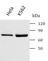
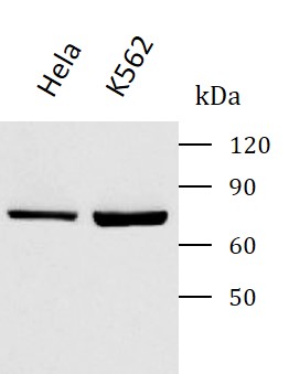
ARG43932 anti-PKC alpha antibody WB image
Western blot: HeLa stained with ARG43932 anti-PKC alpha antibody at 0.5 μg/mL dilution.
-
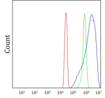
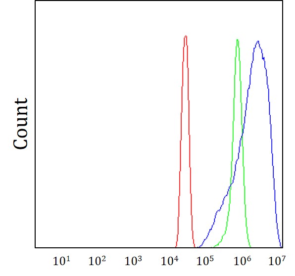
ARG43932 anti-PKC alpha antibody FACS image
Flow Cytometry: SiHa cells stained withARG43932 anti-PKC alpha antibody (blue) at 1 μg/1x10^6 cells dilution.
-
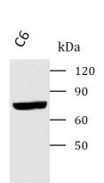
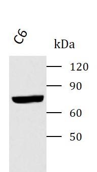
ARG43932 anti-PKC alpha antibody WB image
Western blot: C6 stained with ARG43932 anti-PKC alpha antibody at 0.5 μg/mL dilution.
-
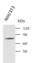
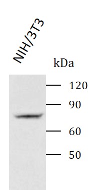
ARG43932 anti-PKC alpha antibody WB image
Western blot: NIH/3T3 stained with ARG43932 anti-PKC alpha antibody at 0.5 μg/mL dilution.