ARG55082
anti-Cellular Apoptosis Susceptibility antibody
anti-Cellular Apoptosis Susceptibility antibody for ICC/IF,IHC-Formalin-fixed paraffin-embedded sections,Western blot and Human,Mouse
Cancer antibody; Cell Biology and Cellular Response antibody; Cell Death antibody; Signaling Transduction antibody
Overview
| Product Description | Rabbit Polyclonal antibody recognizes Cellular Apoptosis Susceptibility |
|---|---|
| Tested Reactivity | Hu, Ms |
| Predict Reactivity | Bov, Zfsh |
| Tested Application | ICC/IF, IHC-P, WB |
| Host | Rabbit |
| Clonality | Polyclonal |
| Isotype | IgG |
| Target Name | Cellular Apoptosis Susceptibility |
| Antigen Species | Human |
| Immunogen | KLH-conjugated synthetic peptide corresponding to aa. 55-84 (C-terminus) of Human Cellular Apoptosis Susceptibility. |
| Conjugation | Un-conjugated |
| Alternate Names | XPO2; Exportin-2; CAS; Exp2; CSE1; Chromosome segregation 1-like protein; Importin-alpha re-exporter; Cellular apoptosis susceptibility protein |
Application Instructions
| Application Suggestion |
|
||||||||
|---|---|---|---|---|---|---|---|---|---|
| Application Note | * The dilutions indicate recommended starting dilutions and the optimal dilutions or concentrations should be determined by the scientist. | ||||||||
| Positive Control | HeLa |
Properties
| Form | Liquid |
|---|---|
| Purification | Purification with Protein G. |
| Buffer | PBS and 0.09% (W/V) Sodium azide |
| Preservative | 0.09% (W/V) Sodium azide |
| Storage Instruction | For continuous use, store undiluted antibody at 2-8°C for up to a week. For long-term storage, aliquot and store at -20°C or below. Storage in frost free freezers is not recommended. Avoid repeated freeze/thaw cycles. Suggest spin the vial prior to opening. The antibody solution should be gently mixed before use. |
| Note | For laboratory research only, not for drug, diagnostic or other use. |
Bioinformation
| Database Links | |
|---|---|
| Gene Symbol | CSE1L |
| Gene Full Name | CSE1 chromosome segregation 1-like (yeast) |
| Background | Proteins that carry a nuclear localization signal (NLS) are transported into the nucleus by the importin-alpha/beta heterodimer. Importin-alpha binds the NLS, while importin-beta mediates translocation through the nuclear pore complex. After translocation, RanGTP binds importin-beta and displaces importin-alpha. Importin-alpha must then be returned to the cytoplasm, leaving the NLS protein behind. The protein encoded by this gene binds strongly to NLS-free importin-alpha, and this binding is released in the cytoplasm by the combined action of RANBP1 and RANGAP1. In addition, the encoded protein may play a role both in apoptosis and in cell proliferation. Alternatively spliced transcript variants have been found for this gene. [provided by RefSeq, Jan 2012] |
| Function | Export receptor for importin-alpha. Mediates importin-alpha re-export from the nucleus to the cytoplasm after import substrates (cargos) have been released into the nucleoplasm. In the nucleus binds cooperatively to importin-alpha and to the GTPase Ran in its active GTP-bound form. Docking of this trimeric complex to the nuclear pore complex (NPC) is mediated through binding to nucleoporins. Upon transit of a nuclear export complex into the cytoplasm, disassembling of the complex and hydrolysis of Ran-GTP to Ran-GDP (induced by RANBP1 and RANGAP1, respectively) cause release of the importin-alpha from the export receptor. CSE1L/XPO2 then return to the nuclear compartment and mediate another round of transport. The directionality of nuclear export is thought to be conferred by an asymmetric distribution of the GTP- and GDP-bound forms of Ran between the cytoplasm and nucleus. [UniProt] |
| Cellular Localization | Cytoplasm. Nucleus. Note=Shuttles between the nucleus and the cytoplasm |
| Research Area | Cancer antibody; Cell Biology and Cellular Response antibody; Cell Death antibody; Signaling Transduction antibody |
| Calculated MW | 110 kDa |
Images (3) Click the Picture to Zoom In
-
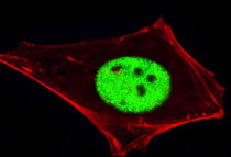
ARG55082 anti-Cellular Apoptosis Susceptibility antibody ICC/IF image
Immunofluorescence: HeLa cells stained with ARG55082 anti-Cellular Apoptosis Susceptibility antibody (green) at 1:100 dilution. Cytoplasmic actin was counterstained with Alexa Fluor® 555 conjugated with Phalloidin (red).
-
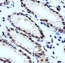
ARG55082 anti-Cellular Apoptosis Susceptibility antibody IHC-P image
Immunohistochemistry: Paraffin-embedded Human stomach tissue stained with ARG55082 anti-Cellular Apoptosis Susceptibility antibody at 1:100 dilution.
-
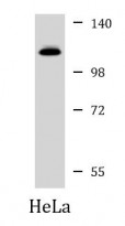
ARG55082 anti-Cellular Apoptosis Susceptibility antibody WB image
Western blot: 35 µg of HeLa cell lysate stained with ARG55082 anti-Cellular Apoptosis Susceptibility antibody at 1:1000 dilution.


