ARG63398
anti-PARD6A / PAR6 antibody
anti-PARD6A / PAR6 antibody for Flow cytometry,IHC-Formalin-fixed paraffin-embedded sections,Western blot and Human
Cancer antibody; Cell Biology and Cellular Response antibody; Developmental Biology antibody; Gene Regulation antibody; Signaling Transduction antibody
Overview
| Product Description | Goat Polyclonal antibody recognizes PARD6A / PAR6 |
|---|---|
| Tested Reactivity | Hu |
| Tested Application | FACS, IHC-P, WB |
| Specificity | This antibody is expected to recognise both reported isoforms (NP_058644.1; NP_001032358.1). |
| Host | Goat |
| Clonality | Polyclonal |
| Isotype | IgG |
| Target Name | PARD6A / PAR6 |
| Antigen Species | Human |
| Immunogen | C-GSRIRGDGSGFSL |
| Conjugation | Un-conjugated |
| Alternate Names | Tax interaction protein 40; TIP-40; PAR-6 alpha; PAR6; PAR6alpha; PAR-6A; PAR-6; PAR6C; Partitioning defective 6 homolog alpha; TAX40 |
Application Instructions
| Application Suggestion |
|
||||||||
|---|---|---|---|---|---|---|---|---|---|
| Application Note | IHC-P: Antigen Retrieval: Microwaved tissue section in Tris/EDTA buffer (pH 9.0). WB: Recommend incubate at RT for 1h. * The dilutions indicate recommended starting dilutions and the optimal dilutions or concentrations should be determined by the scientist. |
Properties
| Form | Liquid |
|---|---|
| Purification | Purified from goat serum by antigen affinity chromatography. |
| Buffer | Tris saline (pH 7.3), 0.02% Sodium azide and 0.5% BSA. |
| Preservative | 0.02% Sodium azide |
| Stabilizer | 0.5% BSA |
| Concentration | 0.5 mg/ml |
| Storage Instruction | For continuous use, store undiluted antibody at 2-8°C for up to a week. For long-term storage, aliquot and store at -20°C or below. Storage in frost free freezers is not recommended. Avoid repeated freeze/thaw cycles. Suggest spin the vial prior to opening. The antibody solution should be gently mixed before use. |
| Note | For laboratory research only, not for drug, diagnostic or other use. |
Bioinformation
| Database Links |
Swiss-port # Q9NPB6 Human Partitioning defective 6 homolog alpha |
|---|---|
| Background | This gene is a member of the PAR6 family and encodes a protein with a PSD95/Discs-large/ZO1 (PDZ) domain and a semi-Cdc42/Rac interactive binding (CRIB) domain. This cell membrane protein is involved in asymmetrical cell division and cell polarization processes as a member of a multi-protein complex. The protein also has a role in the epithelial-to-mesenchymal transition (EMT) that characterizes the invasive phenotype associated with metastatic carcinomas. Alternate transcriptional splice variants, encoding different isoforms, have been characterized. [provided by RefSeq, Jul 2008] |
| Research Area | Cancer antibody; Cell Biology and Cellular Response antibody; Developmental Biology antibody; Gene Regulation antibody; Signaling Transduction antibody |
| Calculated MW | 37 kDa |
| PTM | Phosphorylated by the TGF-beta receptor. |
Images (4) Click the Picture to Zoom In
-
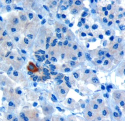
ARG63398 anti-PARD6A / PAR6 antibody IHC-P image
Immunohistochemistry: Paraffin embedded Human Pancreas. (Microwaved antigen retrieval with Tris/EDTA buffer pH9) stained with ARG63398 anti-PARD6A / PAR6 antibody at 10 µg/ml dilution followed by HRP-staining.
-
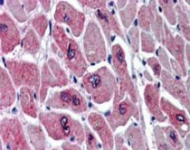
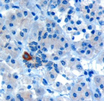
ARG63398 anti-PARD6A / PAR6 antibody IHC-P image
Immunohistochemistry: Paraffin embedded Human Heart. (Steamed antigen retrieval with citrate buffer pH 6) stained with ARG63398 anti-PARD6A / PAR6 antibody at 5 µg/ml dilution followed by AP-staining.
-
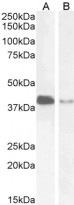
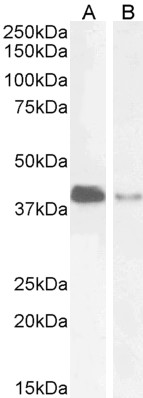
ARG63398 anti-PARD6A / PAR6 antibody WB image
Western blot: 35 µg of Jurkat (A) and U251 (B) cell lysates (in RIPA buffer) stained with ARG63398 anti-PARD6A / PAR6 antibody at 2 µg/ml dilution and incubated at RT for 1 hour.
-

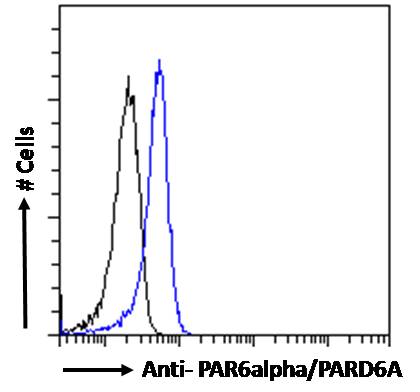
ARG63398 anti-PARD6A / PAR6 antibody FACS image
Flow Cytometry: Paraformaldehyde-fixed Jurkat cells permeabilized with 0.5% Triton. Cells were stained with ARG63398 anti-PARD6A / PAR6 antibody (blue line) at 10 µg/ml dilution for 1 hour, followed by incubation with Alexa FluorR 488 labelled secondary antibody. IgG control: Unimmunized goat IgG (black line).