ARG42962
anti-RRBP1 antibody
anti-RRBP1 antibody for Flow cytometry,ICC/IF,IHC-Formalin-fixed paraffin-embedded sections,Western blot and Human
Overview
| Product Description | Rabbit Polyclonal antibody recognizes RRBP1 |
|---|---|
| Tested Reactivity | Hu |
| Tested Application | FACS, ICC/IF, IHC-P, WB |
| Host | Rabbit |
| Clonality | Polyclonal |
| Isotype | IgG |
| Target Name | RRBP1 |
| Antigen Species | Human |
| Immunogen | Synthetic peptide corresponding to a sequence of Human RRBP1. (EKEKKLTSDLGRAATRLQELLKTTQEQLAREKDTVKKLQEQLEKAED) |
| Conjugation | Un-conjugated |
| Alternate Names | Ribosome-binding protein 1; ES130; Ribosome receptor protein; hES; ES/130-related protein; ES/130; 180 kDa ribosome receptor homolog; RRp |
Application Instructions
| Application Suggestion |
|
||||||||||
|---|---|---|---|---|---|---|---|---|---|---|---|
| Application Note | IHC-P: Antigen Retrieval: Heat mediation was performed in Citrate buffer (pH 6.0) for 20 min. * The dilutions indicate recommended starting dilutions and the optimal dilutions or concentrations should be determined by the scientist. |
||||||||||
| Observed Size | ~ 190 kDa |
Properties
| Form | Liquid |
|---|---|
| Purification | Affinity purification with immunogen. |
| Buffer | 0.2% Na2HPO4, 0.9% NaCl, 0.05% Sodium azide and 4% Trehalose. |
| Preservative | 0.05% Sodium azide |
| Stabilizer | 4% Trehalose |
| Concentration | 0.5 mg/ml |
| Storage Instruction | For continuous use, store undiluted antibody at 2-8°C for up to a week. For long-term storage, aliquot and store at -20°C or below. Storage in frost free freezers is not recommended. Avoid repeated freeze/thaw cycles. Suggest spin the vial prior to opening. The antibody solution should be gently mixed before use. |
| Note | For laboratory research only, not for drug, diagnostic or other use. |
Bioinformation
| Database Links | |
|---|---|
| Gene Symbol | RRBP1 |
| Gene Full Name | ribosome binding protein 1 |
| Background | This gene encodes a ribosome-binding protein of the endoplasmic reticulum (ER) membrane. Studies suggest that this gene plays a role in ER proliferation, secretory pathways and secretory cell differentiation, and mediation of ER-microtubule interactions. Alternative splicing has been observed and protein isoforms are characterized by regions of N-terminal decapeptide and C-terminal heptad repeats. Splicing of the tandem repeats results in variations in ribosome-binding affinity and secretory function. The full-length nature of variants which differ in repeat length has not been determined. Pseudogenes of this gene have been identified on chromosomes 3 and 7, and RRBP1 has been excluded as a candidate gene in the cause of Alagille syndrome, the result of a mutation in a nearby gene on chromosome 20p12. [provided by RefSeq, Apr 2012] |
| Function | Acts as a ribosome receptor and mediates interaction between the ribosome and the endoplasmic reticulum membrane. [UniProt] |
| Cellular Localization | Endoplasmic reticulum membrane; Single-pass type III membrane protein. [UniProt] |
| Calculated MW | 152 kDa |
Images (8) Click the Picture to Zoom In
-
ARG42962 anti-RRBP1 antibody ICC/IF image
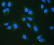
Immunofluorescence: U2OS cells were blocked with 10% goat serum and then stained with ARG42962 anti-RRBP1 antibody (green) at 2 µg/ml dilution, overnight at 4°C. DAPI (blue) for nuclear staining.
-
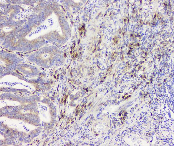
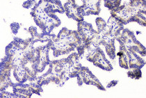
ARG42962 anti-RRBP1 antibody IHC-P image
Immunohistochemistry: Paraffin-embedded Human intestinal cancer tissue. Antigen Retrieval: Heat mediation was performed in Citrate buffer (pH 6.0) for 20 min. The tissue section was blocked with 10% goat serum. The tissue section was then stained with ARG42962 anti-RRBP1 antibody at 1 µg/ml dilution, overnight at 4°C.
-
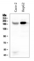
ARG42962 anti-RRBP1 antibody WB image
Western blot: 50 µg of sample under reducing conditions. Caco-2 and HepG2 whole cell lysates stained with ARG42962 anti-RRBP1 antibody at 0.5 µg/ml dilution, overnight at 4°C.
-
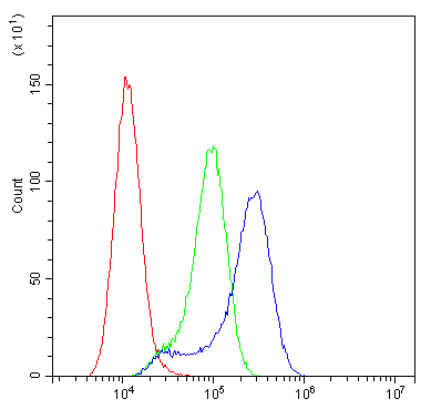
ARG42962 anti-RRBP1 antibody FACS image
Flow Cytometry: A549 cells were blocked with 10% normal goat serum and then stained with ARG42962 anti-RRBP1 antibody (blue) at 1 µg/10^6 cells for 30 min at 20°C, followed by incubation with DyLight®488 labelled secondary antibody. Isotype control antibody (green) was Rabbit IgG (1 µg/10^6 cells) used under the same conditions. Unlabelled sample (red) was also used as a control.
-
ARG42962 anti-RRBP1 antibody IHC-P image
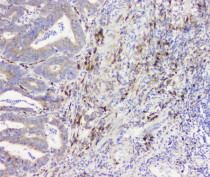
Immunohistochemistry: Paraffin-embedded Human intestinal cancer tissue. Antigen Retrieval: Heat mediation was performed in Citrate buffer (pH 6.0) for 20 min. The tissue section was blocked with 10% goat serum. The tissue section was then stained with ARG42962 anti-RRBP1 antibody at 1 µg/ml dilution, overnight at 4°C.
-
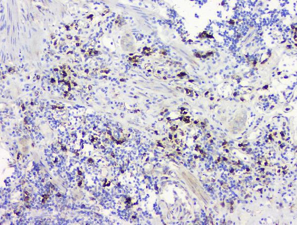
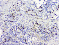
ARG42962 anti-RRBP1 antibody IHC-P image
Immunohistochemistry: Paraffin-embedded Human thyroid cancer tissue. Antigen Retrieval: Heat mediation was performed in Citrate buffer (pH 6.0) for 20 min. The tissue section was blocked with 10% goat serum. The tissue section was then stained with ARG42962 anti-RRBP1 antibody at 1 µg/ml dilution, overnight at 4°C.
-
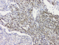
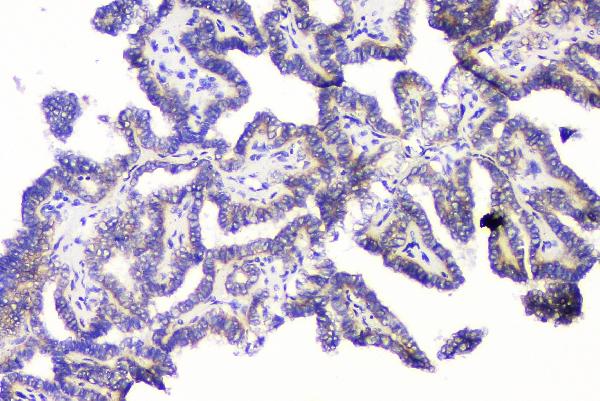
ARG42962 anti-RRBP1 antibody IHC-P image
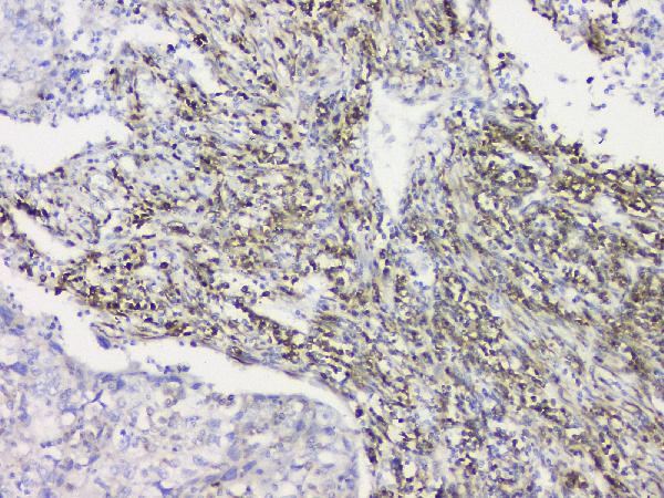
Immunohistochemistry: Paraffin-embedded Human lung cancer tissue. Antigen Retrieval: Heat mediation was performed in Citrate buffer (pH 6.0) for 20 min. The tissue section was blocked with 10% goat serum. The tissue section was then stained with ARG42962 anti-RRBP1 antibody at 1 µg/ml dilution, overnight at 4°C.
-
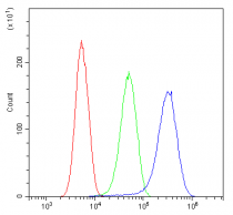
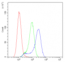
ARG42962 anti-RRBP1 antibody FACS image
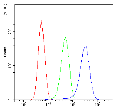
Flow Cytometry: SiHa cells were blocked with 10% normal goat serum and then stained with ARG42962 anti-RRBP1 antibody (blue) at 1 µg/10^6 cells for 30 min at 20°C, followed by incubation with DyLight®488 labelled secondary antibody. Isotype control antibody (green) was Rabbit IgG (1 µg/10^6 cells) used under the same conditions. Unlabelled sample (red) was also used as a control.